

![NRF2 antibody [N2C2], Internal产品图](https://img1.dxycdn.com/2022/0328/677/1261213704005200453-14.jpg)
- ¥1700 - 4000
- GeneTex

- 美国
- GTX103322
- 2025年07月15日
- WB, ICC/IF, IHC-P, FACS, IP, ChIP assay
- Rabbit
- Human, Mouse, Rat, Zebrafish, Alligator, Plant, Bird

企业认证
相关产品推荐更多 >
万千商家帮你免费找货
0 人在求购买到急需产品
相关阅读
源叶生物 十万种现货试剂速发,品质稳、品类全,实验不再等!Takara 秋促来袭,提取+反转录+定量套装仅 1200 元abinScience 体内级抗体 全年锁价 低至 ¥1200阿拉丁 22万+ 科研级产品现货速发,品质严控、科研无忧- 详细信息
- 文献和实验
- 技术资料
- 免疫原:
Recombinant protein encompassing a sequence within the center region of human NRF2. The exact sequence is proprietary.
- 亚型:
IgG
- 形态:
Liquid
- 保存条件:
Store as concentrated solution. Centrifuge briefly prior to opening vial. For short-term storage (1-2 weeks), store at 4ºC. For long-term storage, aliquot and store at -20ºC or below. Avoid multiple freeze-thaw cycles.
- 克隆性:
Polyclonal
- 标记物:
Unconjugated
- 适应物种:
Human, Mouse, Rat, Zebrafish, Alligator, Plant, Bird
- 保质期:
12 months from the shipping date of the product.
- 抗原来源:
Human
- 目录编号:
GTX103322
- 级别:
Primary Antibodies
- 库存:
Available
- 供应商:
GeneTex
- 宿主:
Rabbit
- 应用范围:
WB, ICC/IF, IHC-P, FACS, IP, ChIP assay
- 浓度:
0.41 mg/ml (Please refer to the vial label for the specific concentration.)
- 靶点:
NRF2
- 抗体英文名:
NRF2 antibody [N2C2], Internal
- 抗体名:
NRF2 抗体 [N2C2], Internal
- 规格:
100 μl/25 μl
| 规格: | 100 μl | 产品价格: | ¥4000.0 |
|---|---|---|---|
| 规格: | 25 μl | 产品价格: | ¥1700.0 |
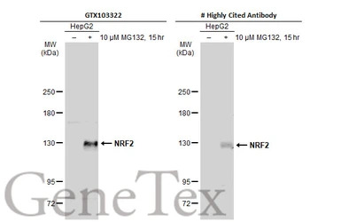
Untreated (–) and treated (+) HepG2 whole cell extracts (30 μg) were separated by 5% SDS-PAGE, and the membranes were blotted with NRF2 antibody [N2C2], Internal (GTX103322) diluted at 1:500 and competitor's antibody diluted at 1:500. The HRP-conjugated anti-rabbit IgG antibody (GTX213110-01) was used to detect the primary antibody.
*The competitor is not affiliated with GeneTex and does not endorse this product.
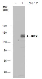
NRF2 antibody detects NRF2 protein by western blot analysis. Non-transfected (-) and NRF2-transfected (+, including 3xFlag-tag) 293T whole cell extracts (30 μg) were separated by 5% SDS-PAGE, and the membrane was blotted with NRF2 antibody (GTX103322) diluted by 1:1000. The HRP-conjugated anti-rabbit IgG antibody (GTX213110-01) was used to detect the primary antibody.
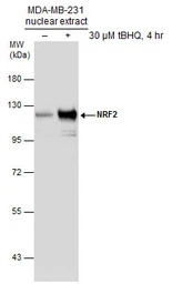
Untreated (–) and treated (+) MDA-MB-231 nuclear extracts (30 μg) were separated by 7.5% SDS-PAGE, and the membrane was blotted with NRF2 antibody [N2C2], Internal (GTX103322) diluted at 1:1000.
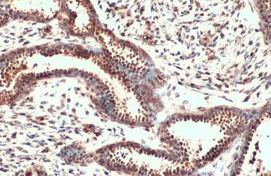
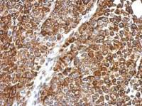
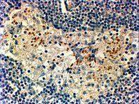
NRF2 antibody [N2C2], Internal detects NRF2 protein at cytoplasm and nucleus by immunohistochemical analysis.
Sample: Paraffin-embedded human breast carcinoma.
NRF2 stained by NRF2 antibody [N2C2], Internal (GTX103322) diluted at 1:500.
Antigen Retrieval: Citrate buffer, pH 6.0, 15 min
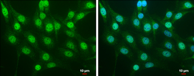
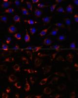
NRF2 antibody [N2C2], Internal detects NRF2 protein at cytoplasm and nucleus by immunofluorescent analysis.
Sample: NIH/3T3 cells were fixed in 4% PFA at RT for 15 min.
Green: NRF2 protein stained by NRF2 antibody [N2C2], Internal (GTX103322) diluted at 1:500.
Blue: Hoechst 33342 staining.
Scale bar = 10 μm.
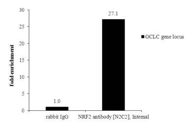
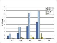
ChIP was performed with HepG2 chromatin extract and 5 μg of either normal rabbit IgG or anti-NRF2 antibody. The precipitated DNA was detected by PCR with primer set targeting to GCLC gene locus.
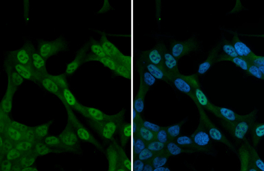
NRF2 antibody [N2C2], Internal detects NRF2 protein at nucleus by immunofluorescent analysis.
Sample: Neuro2A cells were fixed in 4% PFA at RT for 15 min.
Green: NRF2 stained by NRF2 antibody [N2C2], Internal (GTX103322) diluted at 1:1000.
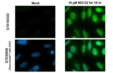
NRF2 antibody [N2C2], Internal detects NRF2 protein at nucleus by immunofluorescent analysis.
Sample: Mock and treated HeLa cells were fixed in 4% PFA at RT for 15 min.
Green: NRF2 stained by NRF2 antibody [N2C2], Internal (GTX103322) diluted at 1:500.
Blue: Fluoroshield with DAPI (GTX30920).
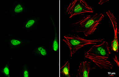
NRF2 antibody [N2C2], Internal detects NRF2 protein at nucleus by immunofluorescent analysis.
Sample: HeLa cells were fixed in 4% PFA at RT for 15 min.
Green: NRF2 stained by NRF2 antibody [N2C2], Internal (GTX103322) diluted at 1:1000.
Red: phalloidin, a cytoskeleton marker, diluted at 1:200.
Scale bar= 10 μm.
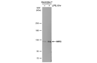
Untreated (–) and treated (+) RAW264.7 whole cell extracts (30 μg) were separated by 5% SDS-PAGE, and the membrane was blotted with NRF2 antibody [N2C2], Internal (GTX103322) diluted at 1:500. The HRP-conjugated anti-rabbit IgG antibody (GTX213110-01) was used to detect the primary antibody.
风险提示:丁香通仅作为第三方平台,为商家信息发布提供平台空间。用户咨询产品时请注意保护个人信息及财产安全,合理判断,谨慎选购商品,商家和用户对交易行为负责。对于医疗器械类产品,请先查证核实企业经营资质和医疗器械产品注册证情况。
 文献和实验
文献和实验Preparation and Assay of Acetylcholinesterase Antibody
structures mimicking external antigen results from the fact that the conformation of some of the anti-idiotypic antibodies may be complementary to the three-dimensional structure of the binding site of the first antibody, and therefore, represents
Protocol for anti-HA antibody Western Blotting
film for ~2 mins. Notes on the primary antibody: Source : Covance/Babco Description: Crude ascites fluid monoclonal anti-HA antibody (CATLN#135032002, 11mms-101R, 0.5 ml) Handling: Aliquoted into 10ul portions in -20℃ Internal notes: labelled anti
Analysis of Antibody-Antigen Interactions Using Surface Plasmon Resonance
Accurate analyses of antibody specificities and affinities are critical to understanding their role in antibody-mediated neutralization of HIV-1 in vitro and their potential activity against HIV-1 in vivo. Multiple immunologic tools
 技术资料
技术资料暂无技术资料 索取技术资料

