

- ¥5400
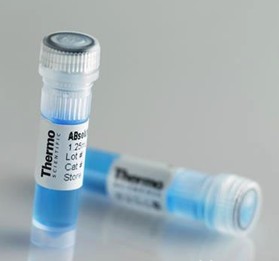
- thermo
- L7012
- USA
- 2026年01月22日

企业认证
相关产品推荐更多 >

万千商家帮你免费找货
0 人在求购买到急需产品
- 详细信息
- 询价记录
- 文献和实验
- 技术资料
- 供应商:
L7012
- 库存:
大量
- 英文名:
LIVE/DEAD™ BacLight™
- 保质期:
一年
- 保存条件:
低温
- 规格:
1 kit
Invitrogen™
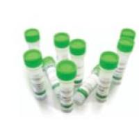
LIVE/DEAD™ BacLight™ Bacterial Viability Kit, for microscopy & quantitative assays
描述
The LIVE/DEAD BacLight Bacterial Viability Kit *for microscopy and quantitative assays* is a convenient and easy-to-use kit for monitoring the viability of bacterial populations as a function of the membrane integrity of the cell. Cells with a compromised membrane that are considered to be dead or dying will stain red, whereas cells with an intact membrane will stain green.规格
| Detection Method: | Fluorescent |
|---|---|
| Compatible Cells: | Bacteria |
| Flow Cytometer Laser Lines: | 488 |
| Excitation/Emission: | SYTO® 9: 485⁄498, PI: 535⁄617 |
| 外形: | Solution |
| 适用于(设备): | Fluorescence Microscope, Fluorometer, Flow Cytometer, Microplate Reader |
| Label or Dye: | SYTO® 9, propidium iodide |
| 形式: | Tube(s), Slide(s) |
| Label Type: | Other Label(s) or Dye(s) |
| 页反应的: | flow cytometry: 200, 96-well plates: ≥ 1,000 |
| Shipping Condition: | Room Temperature |
| Solubility: | DMSO (Dimethylsulfoxide) |
| Product Line: | BacLight™, LIVE/DEAD® |
内容及储存
Store in freezer (-5 to -30°C) and protect from light.
风险提示:丁香通仅作为第三方平台,为商家信息发布提供平台空间。用户咨询产品时请注意保护个人信息及财产安全,合理判断,谨慎选购商品,商家和用户对交易行为负责。对于医疗器械类产品,请先查证核实企业经营资质和医疗器械产品注册证情况。
- 作者
- 内容
- 询问日期
 文献和实验
文献和实验Dendritic cells (DC) are unique in their ability to initiate a primary immune response by the presentation of soluble Ags to T cells. Recent studies have shown that DC also phagocytose particulate Ags including the intracellular pathogen Mycobacterium tuberculosis. However, it is not known whether DC contain the growth of intracellular organisms or allow unlimited replication. To address this question, we infected human DC with a virulent strain of M. tuberculosis and monitored the intracellular growth. The bacteria grew two orders of magnitude within 7 days of culture. Among cytokines known to modulate mycobacterial growth particularly in murine macrophages (TNF-alpha, IFN-gamma, TGF-beta, IL-4), only IL-10 modulated the growth in human DC. This effect was specific for immature dendritic cells, as IL-10 did not induce growth inhibition in human macrophages. In searching for the mechanism of growth inhibition, we found that IL-10 induces the down-regulation of the DC marker CD1, while the macrophage marker CD14 was up-regulated. Functionally, IL-10-treated cells had a reduced capacity to induce an alloresponse, but phagocytic uptake of M. tuberculosis was more efficient. We also show that DC are inferior to macrophages in containing mycobacterial growth. These findings show that IL-10 converts DC into macrophage-like cells, thereby inducing the growth inhibition of an intracellular pathogen. At the site of a local immune response, such as a tuberculous granuloma, IL-10 might therefore participate in the composition of the cellular microenvironment by affecting the maturity and function of DC.
AIMS: The aim of this study was to use confocal laser scanning microscopy (CLSM) to examine the spatial distribution of both viable and nonviable bacteria within microcosm dental plaques grown in vitro. Previous in vivo studies have reported upon the distribution of viable bacteria only. METHODS AND RESULTS: Oral biofilms were grown on hydroxyapatite (HA) discs in a constant-depth film fermenter (CDFF) from a saliva inoculum. The biofilms were stained with the BacLight LIVE/DEAD system and examined by CLSM. Fluorescence intensity profiles through the depth of the biofilm showed an offset between the maximum viable intensity and the maximum nonviable intensity. Topographical differences between the surface properties of the viable and nonviable biofilm virtual surfaces were also measured. CONCLUSIONS: The profile of fluorescence intensity from viable and nonviable staining suggested that the upper layers of the biofilm contain proportionally more viable bacteria than the lower regions of the biofilm. SIGNIFICANCE AND IMPACT OF STUDY: Viability profiling records the transition from predominantly viable to nonviable bacteria through biofilms suggesting that this technique may be of use for quantifying the effects of antimicrobial compounds upon biofilms. The distribution of viable bacteria was similar to that found in dental plaque in vivo suggesting that the CDFF produces in vitro biofilms which are comparable to their in vivo counterparts in terms of the spatial distribution of viable bacteria.
Survival of Salmonella typhimurium within macrophages is associated with virulence. Most data on the fate of Salmonella during infection of macrophages are derived from viable counts of intracellular bacteria. These counts are a result of a combination of bacterial death and growth within the intracellular population but may not reflect the true levels of either macrophage killing of Salmonella or bacterial growth inside cells. In this study, two independent methods have been used to obtain a more accurate measurement of absolute levels of both death and growth of Salmonella inside macrophages. A purine auxotroph (purD) was used to measure Salmonella death in the absence of bacterial growth and then bacterial growth was measured by supplementing the purD cultures with adenosine. Numbers of dead and live Salmonella were also quantitated using the BacLight staining system, which distinguishes dead from live bacteria. Both methods demonstrate that killing of Salmonella by macrophages is considerably greater than detected using traditional cell counts and that bacterial inactivation occurs throughout the infection period. Salmonella was inactivated at a similar rate in both J774 macrophages (most permissive macrophages) and peritoneal exuadate macrophages (least permissive macrophages), suggesting that the major difference between these cells is the ability to limit bacterial growth. These studies also demonstrate that growth of Salmonella within murine macrophages occurs simultaneously with significant amounts of bacterial death. Identifying the factors responsible for shifting the interaction between macrophages and bacteria toward conditions that favor bacterial growth will be critical to understanding Salmonella virulence.
PCR techniques have significantly improved the detection and identification of bacterial pathogens. Even so, the lack of differentiation between DNA from viable and dead cells is one of the major challenges for diagnostic DNA-based methods. Certain nucleic acid-binding dyes can selectively enter dead bacteria and subsequently be covalently linked to DNA. Ethidium monoazide (EMA) is a DNA intercalating dye that enters bacteria with damaged membranes. This dye can be covalently linked to DNA by photoactivation. Our goal was to utilize the irreversible binding of photoactivated EMA to DNA to inhibit the PCR of DNA from dead bacteria. Quantitative 5'-nuclease PCR assays were used to measure the effect of EMA. The conclusion from the experiments was that EMA covalently bound to DNA inhibited the 5'-nuclease PCR. The maximum inhibition of PCR on pure DNA cross-linked with EMA gave a signal reduction of approximately -4.5 log units relative to untreated DNA. The viable/dead differentiation with the EMA method was evaluated through comparison with BacLight staining (microscopic examination) and plate counts. The EMA and BacLight methods gave corresponding results for all bacteria and conditions tested. Furthermore, we obtained a high correlation between plate counts and the EMA results for bacteria killed with ethanol, benzalkonium chloride (disinfectant), or exposure to 70 degrees C. However, for bacteria exposed to 100 degrees C, the number of viable cells recovered by plating was lower than the detection limit with the EMA method. In conclusion, the EMA method is promising for DNA-based differentiation between viable and dead bacteria.
The dnaA operon of Escherichia coli contains the genes dnaA, dnaN, and recF encoding DnaA, beta clamp of DNA polymerase III holoenzyme, and RecF. When the DnaA concentration is raised, an increase in the number of DNA replication initiation events but a reduction in replication fork velocity occurs. Because DnaA is autoregulated, these results might be due to the inhibition of dnaN and recF expression. To test this, we examined the effects of increasing the intracellular concentrations of DnaA, beta clamp, and RecF, together and separately, on initiation, the rate of fork movement, and cell viability. The increased expression of one or more of the dnaA operon proteins had detrimental effects on the cell, except in the case of RecF expression.
7:点击数据点或对象时,NoviSight 软件可在相应的图像上显示相同的点。 结论 3D 样品的 3D 定量分析看上去很复杂,但是 NoviSight 软件可以从头到尾让您的工作流程得到简化。例如,细胞核识别是微球活/死分析的唯一参数。这种简化工作流程也可应用于相关 3D 模型。
488 nm 激光进行活细胞成像,监测响应激光诱导损伤的 MRE11 募集的动力学过程。 成像条件 物镜:60 倍超级色差校正油浸物镜(PLAPON60XOSC)显微镜:FLUOVIEW FV3000 激光扫描共聚焦显微镜 激光:405 nm(ROI 刺激),488 nm(GFP,绿光) 用于定量测量的温和活细胞成像 由于延时成像过程中激光反复照射产生的光漂白和光毒性问题,会影响实验定量测量的准确性。我们的实验需要在强光刺激与温和活细胞成像之间取得平衡,在激光诱导 DNA 损伤后立即捕获可定
内,大多数细胞器内及细胞质的pH值都在 7.0 左右,然而溶酶体内的 pH 值就比较低,mCherry 的 PK 值比较低,可以用于标记溶酶体内的蛋白。 光稳定性:荧光蛋白被不断激发的过程中,会逐渐失去发光能力。对于高质量成像、荧光信号定量分析以及详细描述生物模型,需要选择光稳定性好的荧光蛋白。在普通荧光成像中,例如激光共聚焦对光稳定性要求不是很高,而超分辨荧光成像对光稳定性要求比较高。 成熟时间:指荧光蛋白翻译后正确折叠并形成发光结构所需的时间。成熟时间较长的荧光蛋白不适合标记短寿命的蛋白,目的
 技术资料
技术资料暂无技术资料 索取技术资料

