

- ¥140
- Cellvis

- D29-10-0-N等
- 2026年04月30日

企业认证
万千商家帮你免费找货
0 人在求购买到急需产品
- 详细信息
- 询价记录
- 文献和实验
- 技术资料
- 库存:
10000
- 现货状态:
大量现货
- 供应商:
杭州欣友生物技术有限公司
- 规格:
10个/包,100个/箱
- 使用进口优质玻片,特别适合激光共聚焦实验
- 使用进口USP class VI无细胞毒性的胶水
- 在无尘车间内生产处理和包装
- 通过细胞生物学相关实验的质量检测
- 无热原(<0.5EU/ml)
- Ɣ射线辐照灭菌
-
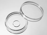
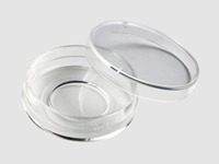
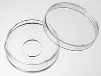
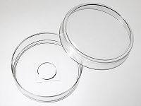
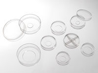
包装规格
产品号 品牌 包装规格 D29-10-0-N Cellvis 底面孔直径10mm,0号玻片,10个每包,100个每箱。 D29-10-1-N Cellvis 底面孔直径10mm,1号玻片,10个每包,100个每箱。 D29-10-1.5-N Cellvis 底面孔直径10mm,1.5号玻片,10个每包,100个每箱。 D29-14-0-N Cellvis 底面孔直径14mm,0号玻片,10个每包,100个每箱。 D29-14-1-N Cellvis 底面孔直径14mm,1号玻片,10个每包,100个每箱。 D29-14-1.5-N Cellvis 底面孔直径14mm,1.5号玻片,10个每包,100个每箱。 D29-20-0-N Cellvis 底面孔直径20mm,0号玻片,10个每包,100个每箱。 D29-20-1-N Cellvis 底面孔直径20mm,1号玻片,10个每包,100个每箱。 D29-20-1.5-N Cellvis 底面孔直径20mm,1.5号玻片,10个每包,100个每箱。 *产品玻片厚度不同,分别是(0号玻片:厚度0.085-0.115mm);(1号玻片:厚度0.13-0.16mm);(1.5号玻片:厚度0.17-0.19mm)。
产品说明
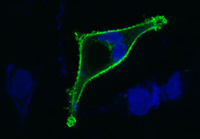
使用Cellvis玻底培养皿
培养Hela细胞的激光共聚焦照片
(感谢军科院陈立勇老师提供本照片)。玻底培养皿主要用于要求放大倍数高、培养器皿底面透光性能好的显微实验,如激光共聚焦显微实验、荧光显微实验和相差显微实验等。玻底培养皿底面薄,透光性能好, 满足了上述实验的要求。同时玻底培养皿底面的小孔也减少了实验过程中抗体等试剂的使用量。
杭州欣友生物技术有限公司在国内生产并为国内生物研究人员提供优质廉价的玻底皿和玻底板。杭州欣友生物技术有限公司生产的玻底皿底面玻璃使用进口的优质玻片,产品采用无细胞毒性的医用胶水粘合,适用于激光共聚焦等需要高分辨率的细胞显微实验,并且能够耐受长期的细胞培养。
我们生产的35mm系列玻底皿使用专门的模具生产,产品在一致性和使用便利上均超过同类进口产品。塑料皿使用高透明度的USP class VI 聚苯乙烯为原料制成。产品表面经过特殊处理,适合贴壁细胞的培养。产品在无尘车间中生产,保证洁净度。
玻底培养皿和玻底培养板使用方法(以35mm皿,10mm孔为例)
- 预平衡:在玻底培养皿加入3ml培养液,在培养箱中放置15分钟。
- 加细胞:吸去培养液,在底孔中加入500ul含细胞的培养液。在培养箱中放置2小时,让细胞沉降贴壁。
- 加培养液:小心加入2-3ml不含细胞的培养液。该步骤用于为细胞提供足够的培养液,同时减少由于水份挥发带来的渗透压的变化。
-
*根据实验的要求,可以合并步骤2和3,在预平衡后直接加入2-3毫升含细胞的培养液。
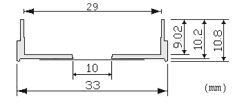
产品尺寸图
风险提示:丁香通仅作为第三方平台,为商家信息发布提供平台空间。用户咨询产品时请注意保护个人信息及财产安全,合理判断,谨慎选购商品,商家和用户对交易行为负责。对于医疗器械类产品,请先查证核实企业经营资质和医疗器械产品注册证情况。
- 作者
- 内容
- 询问日期
 文献和实验
文献和实验- Distinct architecture and composition of mouse axonemal radial spoke head revealed by cryo-EM
Wei Zheng, et al., bioRxiv - Cell Biology 2019
Quote: ... then spread onto laminin-coated 29-mm glass-bottom dishes (Cellvis, D29-14-1.5-N). Then Spermatozoa and mEPCs were pre-extracted with 0.5% Triton X-100 in PBS for 30 secs ... - Distinct architecture and composition of mouse axonemal radial spoke head revealed by cryo-EM
Wei Zheng, et al., bioRxiv - Cell Biology 2019
Quote: ... the cells were transferred to laminin coated 29mm glass bottom dishes (Cellvis, D29-14-1.5-N) and then starved in serum free medium to induce differentiation into ependymal cells. - Organized cannabinoid receptor distribution in neurons revealed by super-resolution fluorescence imaging
Hui Li, et al., bioRxiv - Neuroscience 2020
Quote: ... Fisher Brand) or 29mm Glass (#1.5 cover glass) bottom dishes (D29-20-1.5-N, Cellvis). Neuronal cultures were maintained in the culture medium in a humidified atmosphere with 5% CO2 at 37°C ... - PP2A forestalls mitophagy by dephosphorylating Parkin and ubiquitin
Shang-Xiang Ye, et al., bioRxiv - Biochemistry 2022
Quote: ... coated glass-bottom dishes (Cellvis, catalog number D29-20-1-N) for live-cell imaging ... - Fear extinction is regulated by long noncoding RNA activity at the synapse
Wei-Siang Liau, et al., bioRxiv - Neuroscience 2022
Quote: Primary cortical neurons were cultured and imaged on a glass bottom dish (Cellvis D29-20-1.5-N) as reported previously 60 ... - Endometrial decidualization status modulates endometrial perivascular complexity and trophoblast outgrowth in gelatin hydrogels
Samantha G. Zambuto, et al., bioRxiv - Bioengineering 2022
Quote: Hydrogels were imaged using glass bottom confocal (In Vitro Scientific, D29-20-1-N) dishes on a DMi8 Yokogawa W1 spinning disc confocal microscope outfitted with a Hamamatsu EM-CCD digital camera (Leica Microsystems) ... - Purinergic signaling controls spontaneous activity in the auditory system throughout early development
Travis A. Babola, et al., bioRxiv - Neuroscience 2020
Quote: ... Cochlear pieces were transferred onto polycarbonate membrane filters (Sterlitech PCT0213100) in a 14mm bottom well dish with #0 cover glass (In Vitro Scientific, D29-14-0-N) filled with 250 µL media containing L-15 media (Invitrogen ... - Imaging the ultrastructures and dynamics of live erythrocyte membranes at the single-molecule level with a far-red probe on a microfluidic platform
Zhiwei Ye, et al., bioRxiv - Biophysics 2021
Quote: ... The PDMS replica was covalently bound to a cell culture dish (CellVis, D29-20-1-N) after surface activation for 15 s in oxygen plasma (Diener electronic ...
Cited publications before 2019 (34)
-
The effect of substrate bulk stiffness on focal and fibrillar adhesion formation in human abdominal aortic endothelial cells
H Hassanisaber, et al., Materials Science and Engineering: C Available online 29 December 2018
Quote: "Prior to imaging, the samples were washed three times with 1× PBS and mounted on 29 mm glass-bottom petri-dishes (Cellvis, D29-20-0-N, California, USA) on 100 μl of 50% (v/v) Glycerol (Thermo Fisher Scientific, G33) in 1× PBS" -
Vertebrate Dynein-f depends on Wdr78 for axonemal localization and is essential for ciliary beat
Y Zhang, et al., Journal of Molecular Cell Biology, 28 July 2018
Quote: "The remaining radial glia-enriched cells were further cultured to ∼80% con- fluency (usually 3 days) and then transferred into the wells of laminin-coated 29-mm glass-bottom dishes (Cellvis, D29-14-1.5-N) at a density of 2 × 10 5 cells per well (SS d–3)" -
Mother centrioles are dispensable for deuterosome formation and function during basal body amplification
H Zhao, et al., BioRxiv, July 20, 2018
Quote: "The cells were then transferred into the wells of laminin-coated 29-mm glass-bottom dishes (Cellvis, D29-14-1.5-N) at a density of 2×10⁵ cells per well and were maintained in serum-free medium to induce multiciliate mEPCs" -
Non-canonical Opioid Signaling Inhibits Itch Transmission in the Spinal Cord of Mice
Admire Munanairi, et al., Cell Reports, Volume 23, Issue 3, 17 April 2018, Pages 866-877
Quote: "PKC Translocation Assay: KOR-GRPR HEK293 cells, transiently expressing PKCδ-EGFP or PKCα-EGFP (kindly provided by Dr. Peter M. Blumbergtaken) were seeded in 29-mm glass bottom dishes (In Vitro Scientific)." -
ADAMTS9-Regulated Pericellular Matrix Dynamics Governs Focal Adhesion-Dependent Smooth Muscle Differentiation
Timothy J. Mead, et al., Cell Reports, Volume 23, Issue 2, 10 April 2018
Quote: "Monolayers of HUSMCs were grown and siRNA-transfected. The cells were re-plated on a glass-bottom dish (D-29-10-0-N; Cellvis, Mountain View, CA) and imaged 24 hr later in a heat/CO2-controlled environmental chamber on an inverted Leica SP8 confocal microscope" -
Microtubule-bundling protein Spef1 enables mammalian ciliary central apparatus formation
J Zheng, et al., Journal of Molecular Cell Biology (2018), 1–11
Quote: "The remaining radial glia-enriched cells were further cultured to ∼90% confluency (usually 4 days) and then transferred into the wells of laminin-coated 29-mm glass-bottom dishes (Cellvis, D29-14-1.5-N) at a density of 2 × 10 5 cells per well" -
In Vivo Superresolution Imaging of Neuronal Structure in the Mouse Brain
BE Urban, et al., IEEE Transactions on Biomedical Engineering ( Volume: 65 , Issue: 1 , Jan. 2018 )
Quote: "Before the solution solidified, 1 mL of the mixture was placed in a 29 mm glass bottom dish with a 20 mm bottom well (D29-20-0-N, In Vitro Scientific) and immediately placed in a 4 °C environment to solidify" -
In vivo super-resolution imaging of neuronal structure in the mouse brain
BE Urban, et al., IEEE Transactions on Biomedic, Volume: 65 Issue: 1
Quote: "1 mL of the mixture was placed in a 29 mm glass bottom dish with a 20 mm bottom well (D29-20-0-N, In Vitro Scientific)" -
Effect of pH on polyethylene glycol (PEG)-induced silk microsphere formation for drug delivery
J Wu, et al., Materials Science and Engineering: C Volume 80, 1 November 2017, Pages 549-55
Quote: "Drug-loaded silk microspheres were suspended in 20 μl ultrapure water which was added to a well of cell culture dish with a glass bottom (29 mm dish with 20 mm bottom well, In Vitro Scientific" -
Cellular Contraction Can Drive Rapid Epithelial Flows
Dhruv K.Vig, et al., Biophysical Journal, Volume 113, Issue 7, 3 October 2017, Pages 1613-1622
Quote: "silane (3-aminopropyl triethoxysilane) (Sigma-Aldrich) was vapor-deposited onto a 29-mm glass-bottom dish with a 14-mm microwell (In Vitro Scientific)"
1. 观察更多样品 倒置显微镜载物台采用诸如 160×110 mm 尺寸 K 接口的通用底座标准,可以选择用于处理各种样品的载物台插件。 这样可以观察多个培养皿或多个载玻片,可以同时进行实验,节省大量时间。这种方法减少了用于更换标本和设置常规实验的时间。 图1.通过用在显微镜载物台上放三个培养皿取代放置一个培养皿,可以缩短用于实验准备和设置优化的时间,继而缩短了在实验室停留的时间 2. 加快速度 接下来就是优化图像采集速度。通常拖慢图像采集速度的瓶颈是滤色片或荧光镜组的机械切换
')2 Fragment(Invitrogen) 上述试剂均由嘉美生物代买。 实验仪器:激光共聚焦扫描显微镜(LeicaTCS-SP5型) 实验步骤: 将细胞每孔2ml(密度为2*106/L)传至激光共聚焦专用培养皿;接种24h后,采用刺激物处理;25min后培养皿内细胞培养液,PBS冲洗3次,2-4%甲醛固定15min;1×PBS缓冲液(0.01M,pH7.4)洗涤3次,每次5min,滴加非免疫山羊血清(内含0.3%Triton X-100)封闭液,覆盖液面2-3mm,室温放置60min,除去
的HFZ1型,它与FE1型加温蚀刻装置一起安装在HUS5型真空喷镀仪中使用。以上两种类型各有优缺点,专用装置优点在于操作方便,能连续制样,效率高。缺点是价格贵;附件装置价格虽便宜,但不能连续操作,效率低。 四、操作方法 冷冻蚀刻的操作方法按以下步骤进行。 显微图像 1.预处理取新鲜组织块,大小为15~3~5mm,用25%戊二醛固定1~3小时。为防止冰晶形成,用30%甘油生理盐水浸泡8~12小时。 2.冷冻断裂是在冷冻
 技术资料
技术资料暂无技术资料 索取技术资料

