

- ¥140 - 240
- Cellvis

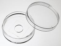
- D35-10-0-N
- 2026年07月12日
企业认证

万千商家帮你免费找货
0 人在求购买到急需产品
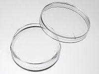
- 详细信息
- 询价记录
- 文献和实验
- 技术资料
- 库存:
1000
- 现货状态:
大量现货
- 供应商:
杭州欣友生物技术有限公司
- 规格:
10个/包,100个/箱
- 使用进口优质玻片,特别适合激光共聚焦实验
- 使用进口USP class VI无细胞毒性的胶水
- 在无尘车间内生产处理和包装
- 通过细胞生物学相关实验的质量检测
- 无热原(<0.5EU/ml)
- Ɣ射线辐照灭菌
-
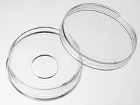
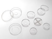
包装规格
产品号 品牌 包装规格 D35-10-0-N Cellvis 底面孔直径10mm,0号玻片,10个每包,100个每箱。 D35-10-1-N Cellvis 底面孔直径10mm,1号玻片,10个每包,100个每箱。 D35-10-1.5-N Cellvis 底面孔直径10mm,1.5号玻片,10个每包,100个每箱。 D35-14-0-N Cellvis 底面孔直径14mm,0号玻片,10个每包,100个每箱。 D35-14-1-N Cellvis 底面孔直径14mm,1号玻片,10个每包,100个每箱。 D35-14-1.5-N Cellvis 底面孔直径14mm,1.5号玻片,10个每包,100个每箱。 D35-20-0-N Cellvis 底面孔直径20mm,0号玻片,10个每包,100个每箱。 D35-20-1-N Cellvis 底面孔直径20mm,1号玻片,10个每包,100个每箱。 D35-20-1.5-N Cellvis 底面孔直径20mm,1.5号玻片,10个每包,100个每箱。 D35-14-1.5GI Cellvis 玻片上有格子,格子在培养皿的里面,细胞养在有格子的这个面上。底面孔直径14mm,1.5号玻片,8个每包,48个每箱。 D35-14-1.5GO Cellvis 玻片上有格子,格子在培养皿的外面,细胞养在玻片没有格子的这个面上。底面孔直径14mm,1.5号玻片,8个每包,48个每箱。 *产品玻片厚度不同,分别是(0号玻片:厚度0.085-0.115mm);(1号玻片:厚度0.13-0.16mm);(1.5号玻片:厚度0.17-0.19mm)。
产品说明
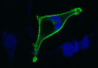
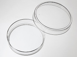
使用Cellvis玻底培养皿
培养Hela细胞的激光共聚焦照片
(感谢军科院陈立勇老师提供本照片)。玻底培养皿主要用于要求放大倍数高、培养器皿底面透光性能好的显微实验,如激光共聚焦显微实验、荧光显微实验和相差显微实验等。玻底培养皿底面薄,透光性能好, 满足了上述实验的要求。同时玻底培养皿底面的小孔也减少了实验过程中抗体等试剂的使用量。
杭州欣友生物技术有限公司在国内生产并为国内生物研究人员提供优质廉价的玻底皿和玻底板。杭州欣友生物技术有限公司生产的玻底皿底面玻璃使用进口的优质玻片,产品采用无细胞毒性的医用胶水粘合,适用于激光共聚焦等需要高分辨率的细胞显微实验,并且能够耐受长期的细胞培养。
我们生产的35mm系列玻底皿使用专门的模具生产,产品在一致性和使用便利上均超过同类进口产品。塑料皿使用高透明度的USP class VI 聚苯乙烯为原料制成。产品表面经过特殊处理,适合贴壁细胞的培养。产品在无尘车间中生产,保证洁净度。
玻底培养皿和玻底培养板使用方法(以35mm皿,10mm孔为例)
- 预平衡:在玻底培养皿加入3ml培养液,在培养箱中放置15分钟。
- 加细胞:吸去培养液,在底孔中加入500ul含细胞的培养液。在培养箱中放置2小时,让细胞沉降贴壁。
- 加培养液:小心加入2-3ml不含细胞的培养液。该步骤用于为细胞提供足够的培养液,同时减少由于水份挥发带来的渗透压的变化。
-
*根据实验的要求,可以合并步骤2和3,在预平衡后直接加入2-3毫升含细胞的培养液。
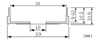
产品尺寸图
风险提示:丁香通仅作为第三方平台,为商家信息发布提供平台空间。用户咨询产品时请注意保护个人信息及财产安全,合理判断,谨慎选购商品,商家和用户对交易行为负责。对于医疗器械类产品,请先查证核实企业经营资质和医疗器械产品注册证情况。
- 作者
- 内容
- 询问日期
 文献和实验
文献和实验- Human photoreceptors switch from autonomous axon extension to cell-mediated process pulling during synaptic marker redistribution
Sarah K. Rempel, et al., bioRxiv - Neuroscience 2021
Quote: ... affixed over a hole in a 35mm petri dish (Falcon #351008) or pre-manufactured dishes (Cellvis #D35-10-1.5-N or #D35-20-1.5-N). Culture dishes were flooded with 2mL RDM (no FBS ... - Mechano-responsiveness of fibrillar adhesions on stiffness-gradient gels
Nuria Barber-Pérez, et al., bioRxiv - Cell Biology 2019
Quote: ... hydrogels were prepared on gridded glass-bottom dishes (Cellvis, D35-14-1.5GO) as above to allow the same area to be located under different microscopes (SDC and AFM). - Thermogenetic control of Ca2+ levels in cells and tissues
Yulia G. Ermakova, et al., bioRxiv - Synthetic Biology 2023
Quote: ... seedlings were transferred to microscope dishes (D35-14-0-N, Cellvis) supplemented with 0.25 MS media ... - An EpCAM/Trop2 mechanostat differentially regulates individual and collective migration of human carcinoma cells
Azam Aslemarz, et al., bioRxiv - Cell Biology 2022
Quote: Glass bottom dishes (Cellvis, D35-20-1.5-N) or 12mm round coverslips were plasma cleaned to create a hydrophilic surface ... - Time-tagged ticker tapes for intracellular recordings
Dingchang Lin, et al., bioRxiv - Neuroscience 2021
Quote: ... or 14 mm glass bottom dishes (CellVis, D35-14-1.5-N) coated with 40 μg/ml poly-L-lysine-coated (P8920 ... - Conformational dynamics regulate SHANK3 actin and Rap1 binding
Siiri I Salomaa, et al., bioRxiv - Cell Biology 2020
Quote: ... 35 mm #1.5 glass-bottom dishes (Cellvis, #D35-14-1.5-N) were coated with bovine plasma fibronectin (Merck-Millipore ... - Calcium flux through ER-TGN contact sites facilitates cargo export
Bulat R. Ramazanov, et al., bioRxiv - Cell Biology 2023
Quote: ... Cells were seeded on Glass bottom dishes (D35-14-1.5-N, Cellvis) at a density 5×10⁴ per dish ... - Negative durotaxis: cell movement toward softer environments
Aleksi Isomursu, et al., bioRxiv - Biophysics 2020
Quote: Glass-bottom dishes (Cellvis, D35-14-1-N) were treated for 20 min at room temperature with 100 μl of Bind-Silane solution – a mixture of 3-(trimethoxysilyl)propylmethacrylate (7.15% by volume ... - Endosomal removal and disposal of dysfunctional, immunostimulatory mitochondrial DNA
Laura E. Newman, et al., bioRxiv - Cell Biology 2022
Quote: ... and mTurquoise-LC3 and plated onto gridded coverslips in 35 mm dishes (Cellvis #D35-14-1.5GI). Cells were stained with PicoGreen and JF635 ... - Spatial regulation of AMPK signaling revealed by a sensitive kinase activity reporter
Danielle L. Schmitt, et al., bioRxiv - Molecular Biology 2021
Quote: ... cells were plated on 35mm glass-bottomed dishes (CellVis D35-14-1.5-N). Cells were transfected 2-24 hours after plating ...
Cited publications before 2019 (161)
-
Induced neural progenitor cells abundantly secrete extracellular vesicles and promote the proliferation of neural progenitors via extracellular signal–regulated kinase pathways
Y Ma, et al., Neurobiology of Disease Volume 124, April 2019, Pages 322-334
Quote: "2.5 × 10 5 WT-NPCs were treated with 15 μg/ml EVs on 35 mm Coverglass-Bottom Dish (Cellvis, #161115) for 24 h" -
The effect of low-intensity ultrasound and met signaling on cellular motility and morphology
N Mazzawi,, Applied Acoustics Volume 143, 1 January 2019, Pages 1-6
Quote: "The fluorescently tagged MDCK cells (pEYFP-mem & pmCherry) were seeded on a glass-coverslip bottomed 35 mm Petri dish (10 mm micro-well #1.5 cover glass, catalog number D35-10-1.5-N, Cellvis), 24 h before experiment" -
Proteomic signature of neuroblastoma cells UKF-NB-4 reveals key role of lysosomal sequestration and the proteasome complex in acquiring chemoresistance to cisplatin
MA Merlos Rodrigo, et al., J. Proteome Res., December 28, 2018
Quote: "Confocal laser scanning microscopy (CLSM) The cells were grown on 35 mm glass bottom culture dishes (In Vitro Scientific, Sunnyvale, CA, USA) for 24 h before treatment." -
A high-avidity biosensor reveals plasma membrane PI(3,4)P2 is predominantly a class I PI3K signaling product
Brady D. Goulden, et al., JCB, December 27, 2018
Quote: "For transfection, cells were seeded in 35-mm tissue culture dishes with 20-mm number 1.5 cover glass apertures (CellVis) coated with 5 µg fibronectin (Life Technologies 33016-015)" -
Muscle-specific stress fibers give rise to sarcomeres in cardiomyocytes
Aidan M Fenix1, et al., ElifeScience, 2018
Quote: "For both live and fixed cell micros-copy, cells were plated and imaged on 35 mm glass bottom dishes with a 10 mm micro-well #1.5 cover glass (Cellvis, Mountain View, CA) coated with 25 mg/mL laminin (114956-81-9, Sigma-Aldrich)." -
Mechanical loading of desmosomes depends on the magnitude and orientation of external stress
Andrew J. Price, et al., Nature Communications volume 9, Article number: 5284 (2018)
Quote: "For imaging experiments, MDCK cell lines were treated with 0.1 μg mL−1 (DPI-ctrl) or 0.5 μg mL−1 (DPI-TS and photometric controls) doxycycline in order to achieve similar levels of construct expression for the cell lines, and plated onto collagen-coated coverslips (Cellvis, D35-20-1.5-N) 48 hours (h) prior to imaging." -
A Litopenaeus vannamei Hemocyanin-Derived Antimicrobial Peptide (Peptide B11) Attenuates Cancer Cells’ Proliferation
Shangjie Liu, et al., Molecules 2018, 23(12), 3202
Quote: "Analysis of Subcellular Localization: HeLa cells (1 × 10⁵) were plated onto a 35-mm glass-bottom dish (In Vitro Scientific) overnight" -
A positive feedback mechanism ensures proper assembly of the functional inner centromere during mitosis in human cells
C Liang, et al., JBC, November 29, 2018
Quote: "Cells expressing H2B-GFP were plated in four-chamber glass-bottomed 35-mm dishes (Cellvis) coated with Poly-D-Lysine, and filmed in a climate-controlled and humidified environment (37°C and 5% CO2). Images were captured every 2 min" -
Optical sensor revealed abnormal nuclease spatial activity on cancer cell membrane
Yongliang Wang, et al., Journal of Biophotonics, 29 November 2018
Quote: "First, 100 µg/ml BSA-biotin (Bovine serum albumin labeled by biotin, A8549, Sigma-Aldrich) and 5 µg/ml fibronectin were incubated on a glass-bottom petri dish (D35-14-1.5-N, In Vitro Scientific) for 30 min." -
Multi-color live-cell super-resolution volume imaging with multi-angle interference microscopy
Youhua Chen, et al., Nature Communications, volume 9, Article number: 4818 (2018)
Quote: "Cells were seeded in 35-mm glass-bottom dishes (Cellvis) and fixed and permeabilized with a –20 °C methanol for 10 min."
View all publications citing "35 mm Glass bottom dishes"
Cellvis (formerly In Vitro Scientific), P.O.Box 390959, Mountain View, CA 940
生长发育的研究,是最为经典而常用的方法之一。1、材料和方法取新生一天的大鼠(wistar种)和小鼠(昆明种)。用眼科剪在无菌条件下除去背部皮肤, 然后剪取一段脊髓,背侧朝上置于灭菌毛玻璃片上,在解剖显微镜下沿椎管两侧水平剪除腹侧一半椎骨,暴露脊髓和神经节,用解剖镊分离出神经节。鸡胚背根神经节的取材方法同前。 剥除神经节被膜, 用0.125%胰蛋白酶消化(37℃ 30min)分散后用种植(Plating)培养液稀释成0.2×10⁵ 个细胞/ml密度的细胞悬液,接种于涂有鼠尾胶的35mm塑料培养皿中,每皿2ml
向胸主动脉注入D-Hank’s液,驱除残血,取主动脉弓至肾动脉一段,两端结扎剪断,放入60℃预热无菌水中2秒。然后置于准备好的RPMi1640培养基(含双抗)中。 2.剪切:在超净台上,将血管置于培养皿中,用眼科镊小心剥离外膜面的结缔组织,并从根部剪去所有肋间动脉。无血清培养基冲洗数遍,去除残血后,将动脉转移至另一含少量培基的无菌培养皿中,用刀片将主动脉两末端切去,剩下的主动脉切成宽1~1.5mm的环。 3.接种:将动脉环竖直放入35mm培养皿(1%明胶4℃预置过夜,用前2h移入CO2培养箱,用前
培养方法 一、器具:1、培养皿(35mm)(或50cm2培养瓶)2、6孔(35mm)培养板、24孔培养板3、滴管4、小青瓶5、100ml、250ml玻璃瓶6、翻口橡皮塞7、烧杯:1000ml、500ml、100ml、50ml8、容量瓶:1000ml、500ml、100ml9、量筒:50ml、100ml、250ml10、玻璃棒11、pH试纸12、滤器(0.22μl,γ射线菌一次性使用)13、注射器:10ml、20ml14、24mm×24mm盖玻片 二、器具处理: 玻璃器皿、塑料器皿,予清洗、泡酸、三蒸水
 技术资料
技术资料暂无技术资料 索取技术资料

