

- ¥750
- 盖宁生物
- 上海
- GN1097
- 2026年05月28日

企业认证

万千商家帮你免费找货
0 人在求购买到急需产品
- 详细信息
- 文献和实验
- 技术资料
- 保存条件:
常温
- 保质期:
三年
- 英文名:
pDsRed2-C1
- 库存:
现货
- 供应商:
上海盖宁生物
| 质粒类型: | 哺乳细胞表达载体 |
|---|---|
| 启动子: | CMV |
| 克隆方法: | 多克隆位点,限制性内切酶 |
| 载体大小: | 4675 bp |
| 5' 测序引物及序列: | DsRed1-C: 5'd[AGCTGGACATCACCTCCCACAACG] |
| 载体标签: | DsRed2 (Nterm) |
| 载体抗性: | Kanamycin (卡那霉素) |
| 筛选标记: | Neomycin (新霉素) |
| 备注: |
Red fluorescent protein tag
|
订购信息
| 产品编号 | 产品名称 | 规格 | 价格 |
|---|---|---|---|
| 1097 | pDsRed2-C1 | 5ug质粒 |
¥1000.00 |
质粒图谱
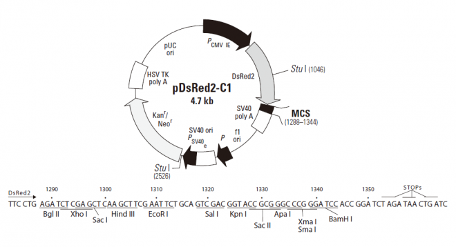
载体描述
pDsRed2-C1 encodes DsRed2, a DsRed variant that has been engineered for faster maturation and lower non-specific aggregation. Derived from the Discosoma sp. red fluorescent protein (drFP583; 1), DsRed2, like its progenitor DsRed1, contains a series of silent base-pair changes that correspond to human codon-usage preferences for high expression in mammalian cells (2). In addition to these changes, DsRed2 contains six amino acid substitutions: V105A, I161T, and S197A, which result in the more rapid appearance of red fluorescence in transfected cell lines; and R2A, K5E, and K9T, which prevent the protein from aggregating. (DsRed2 may, however, form the same tetrameric structure as DsRed1 [3].) In mammalian cell cultures when DsRed2 is expressed constitutively, red-emitting cells can be detected by fluorescence microscopy within 24 hours of transfection. Large insoluble aggregates of protein, often obsrved in bacterial and mammalian cell systems expressing DsRed1, are dramatically reduced in organisms expressing DsRed2. The faster-maturing, more soluble red fluorescent protein is also well tolerated by host cells; mammalian cell cultures transfected with DsRed2 show no obvious signs of reduced viabilityin those cell lines tested, cells expressing DsRed2 display the same morphology (e.g., adherence, light-refraction) and growth characteristics as non-transfected controls.
The multiple cloning site (MCS) in pDsRed2-C1 is positioned between the DsRed2 coding sequence and the SV40 polyadenylation signal (SV40 poly A). Genes cloned into the MCS will be expressed as fusions to the C-terminus of DsRed2 if they are in the same reading frame as DsRed2 and there are no intervening stop codons. A Kozak consensus translation initiation site upstream of DsRed2 increases the translation efficiency in eukaryotic cells (4). SV40 poly A signals downstream of the MCS direct proper processing of the 3' end of mRNA transcripts. The vector backbone also contains an SV40 origin for replication in mammalian cells expressing the SV40 T-antigen, a pUC origin of replication for propagation in E. coli, and an f1 origin for single-stranded DNA production. A neomycin resistance cassette (Neor), consisting of the SV40 early promoter, the neomycin/kanamycin resistance gene of Tn5, and polyadenylation signals from the Herpes simplex virus thymidine kinase (HSV TK) gene, allows stably transfected eukaryotic cells to be selected using G418. A bacterial promoter upstream of this cassette expresses kanamycin resistance in E. coli.
载体应用
pDsRed2-C1 can be used to construct fusions to the C-terminus of DsRed2. If a fusion construct retains the fluorescent properties of the native DsRed2 protein, its expression can be monitored by flow cytometry and its localization in vivo can be determined by fluorescence microscopy. The target gene should be cloned into pDsRed2-C1 so that it is in frame with the DsRed2 coding sequences, with no intervening in-frame stop codons. The recombinant DsRed2 vector can be transfected into mammalian cells using any standard transfection method. If required, stable transformants can be selected using G418 (5). pDsRed2-C1 can also be used as a cotransfection marker; the unmodified vector will express DsRed2.
风险提示:丁香通仅作为第三方平台,为商家信息发布提供平台空间。用户咨询产品时请注意保护个人信息及财产安全,合理判断,谨慎选购商品,商家和用户对交易行为负责。对于医疗器械类产品,请先查证核实企业经营资质和医疗器械产品注册证情况。
 文献和实验
文献和实验*发表【中文论文】请标注:由上海盖宁生物科技有限公司提供;
*发表【英文论文】请标注:From Shanghai Gaining Biotechnology Co., Ltd.
所有酸性液泡。pDsRed2-mito:载体,转染后表达一个融合蛋白(红色荧光蛋白+线粒体基质定位信号),可用来检测线粒体被自噬掉的程度(Mitophagy)。MitoTraker 探针:特异性显示活的线粒体,荧光在经过固定后还能保留。Hsp60:定位与线粒体基质,细胞死亡时不会被释放。Calreticulin(钙网织蛋白):内质网腔。 Note:这些蛋白均为胞浆蛋白,爬片或胰酶消化的细胞在做免疫荧光前需先透膜(permeablize),可采用 0.1% SDS 处理。 参考文献:
pDsredN1 表达 红色 荧光蛋白和目的基因的融合蛋白,目的基因位于N端 kan/neo 4.7kb E. coli/mammals pDsred1-C1 pDsred2-C1 pDsred2C1 表达 红色 荧光蛋白和目的基因的融合蛋白,目的基因位于C端 kan/neo 4.7kb E. coli/mammals pDsRed2-1 pmCherry-C1 pmCherry-C1 表达 红色 荧光蛋白和目的基因的融合蛋白,目的基因位于C端 kan/neo 4.7kb E
啦之类的那一块。这家伙是得了炸药奖的。Clontech公司构建了大量荧光定位质粒,如pEGFP-N1,pDsRed2-N1,pDsRed-Monomer-C1等等。 萤光,英文luminescence,即发光或称生物发光,是指萤火虫荧光素酶,海肾荧光素酶等在底物存在(实际上还需要ATP,氧,辅酶A等,这些试剂盒中已经加入)下发出的光,是生物化学反应,他的实质是酶,没有底物是不能发光的。把LUC利用的最好的是PROMEGA公司,他们有大量试剂盒是基于这两种荧光素酶的,最常见的是检测启动子或增强子活性
 技术资料
技术资料暂无技术资料 索取技术资料

