


- ¥3500 - 4500
- 欣润生物
- 江苏无锡
- IR3006-1
- 2026年03月12日

企业认证
相关产品推荐更多 >


万千商家帮你免费找货
0 人在求购买到急需产品
- 详细信息
- 文献和实验
- 技术资料
- 英文名:
A
- 库存:
10
- 供应商:
欣润生物
- 肿瘤类型:
NO
- 细胞类型:
永生化
- ATCC Number:
11222
- 品系:
SD大鼠
- 组织来源:
主动脉
- 相关疾病:
无
- 物种来源:
大鼠
- 免疫类型:
不详
- 细胞形态:
梭形
- 是否是肿瘤细胞:
否
- 器官来源:
血管
- 运输方式:
常温
- 年限:
5年
- 生长状态:
贴壁生长
- 规格:
T25方瓶
产品介绍
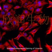
细胞名称:大鼠主动脉内皮细胞系
背景描述:大鼠主动脉内皮细胞系来源于成年大鼠主动脉组织,大鼠主动脉内皮细胞系是组成主动脉腔面单层扁平上皮样内皮细胞,大多呈梭形,人主动脉内皮细胞系所产生和分泌的生物活性物质对维持血管张力,调节血压,抗血栓形成等有重要作用,在高血压,心、脑血管疾病的发病机制中有重要病理生理学意义。。
产品货号:IR3006
细胞类型:永生化细胞
传代能力:可传代30代左右
细胞形态:梭形
完全培养基:大鼠主动脉内皮细胞系完全培养基
支原体检测:阴性
培养条件:37℃,5%CO2
发货方式:T25方瓶
货期:1周左右
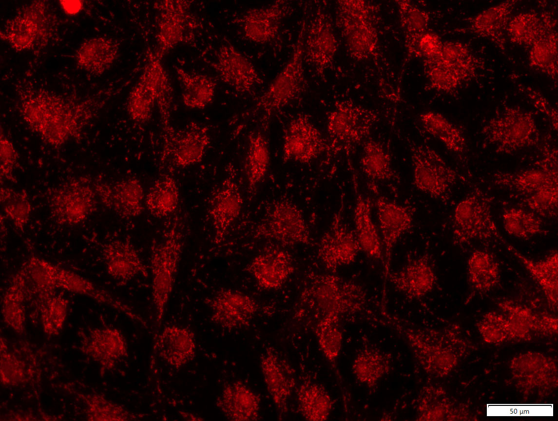
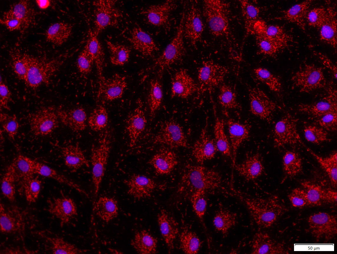
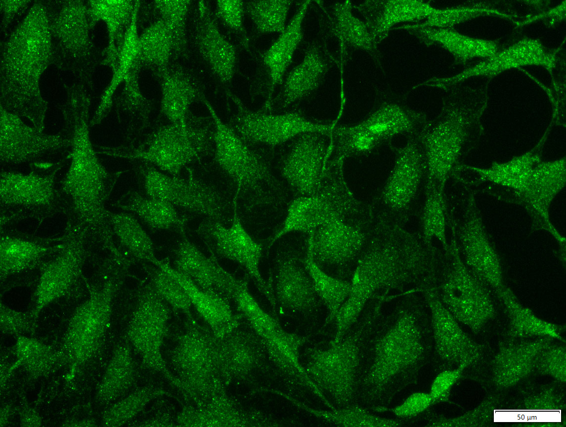
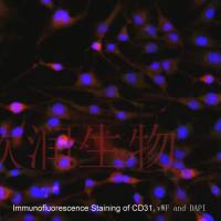
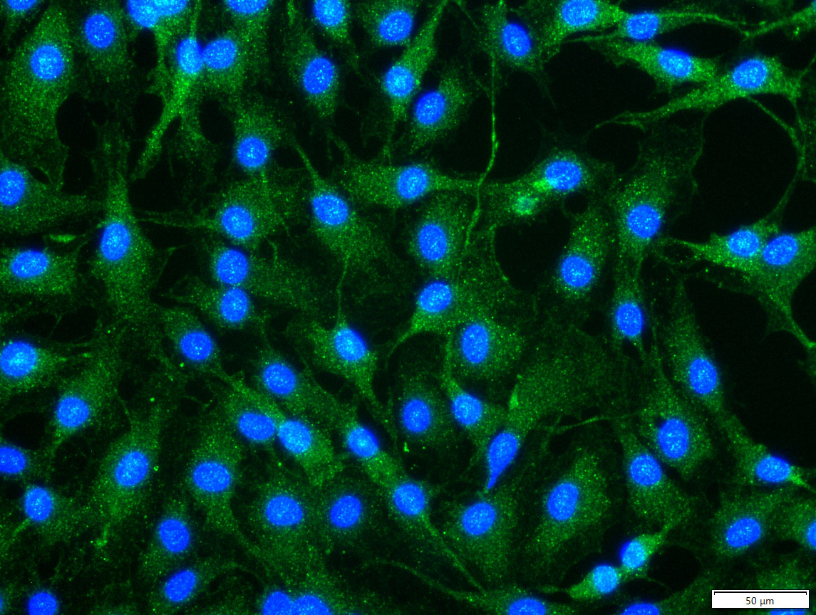
vWF和CD31免疫荧光检测呈阳性


Isolation and characterization of immortalized porcine aortic endothelial cell lines
Primary porcine endothelial cells have a limited life span in culture. After four to five passages, they tend to de-differentiate and eventually reach senescence. The aim of this work was to establish immortalized porcine aortic endothelial cell lines (AOCs) to facilitate in vitro studies of different pathological process involving the endothelium. Primary porcine aortic endothelial cells (PAECs) were transfected with a plasmid containing the SV40 genome and selected on the basis of morphological and phenotypical features. Flow cytometry analysis demonstrated uptake of acetylated low density lipoproteins (Ac-LDL) and constitutive expression of SLA class I, CD29, CD31, CD41/61, CD80/86, CD46, SWC3, and LAMP-1 antigens by all analyzed lines and showed little differences to primary cells. The functional similarity between primary and immortalized endothelial cells was demonstrated in a cytotoxicity assay using a human natural killer cell line (NKL) as effector. The AOCs cell lines should be valuable tools for in vitro study of the human immune response against pig endothelial cells.
风险提示:丁香通仅作为第三方平台,为商家信息发布提供平台空间。用户咨询产品时请注意保护个人信息及财产安全,合理判断,谨慎选购商品,商家和用户对交易行为负责。对于医疗器械类产品,请先查证核实企业经营资质和医疗器械产品注册证情况。
 文献和实验
文献和实验Chicken intestinal epithelial cells were obtained from NEWGAINBIO company. Cells were cultured on 37℃, with 5% CO2, in the Ham’s F-12 Nutrient (DMEM/12) that contained the following supplementations: fetal bovine serum (5%), in-sulin (5 µg/mL), transferrin (5 µg/mL), selenium (5 ng/mL), epidermal growth factor (5 ng/mL) and penicillin-streptomycin (100–100 U/mL) for cell culturing (full DMEM/12). Experiments were performed with chicken intestinal epithelial cells and working solutions were prepared with plain DMEM/12 without supplementation. For the investigations, cells were seeded onto 96-well, 24-well or 6-well polystyrene cell culture plates.
Primary hVICs (passage 2) were cultured to 50–60% confluence and infected with pGMLV-SV40T-puro lentivirus (NewgainBio, Wuxi, China) at a multiplicity of infection of 80 supplemented with 5 µg/mL polybrene (Sigma-Aldrich, Buchs, Switzerland).
Tissue was cultured until cells became visible around the tissue, and when the fusion reached 90% (FIGURE 1A) §ask ¦lled with the prepared culturing medium was sent to the company for further immortalisation. Cell immortalisation was done for cell stability and longer-term use. Immortalised cells were cultured with 10% FBS and 1% PS in the DMEM medium. After the cells multiplied and merged, they were routinely passed and grown ( NEWGAINBIO Inc. Wuxi, Jiangsu, China) (FIGURE 1B-C).
Mouse primary cultured renal vascular ECs and VSMCs were obtained from Newgainbio company, which were tested by Factor VIII and α-smooth muscle actin (α-SMA), the marker of ECs and VSMCs. RNeasy Mini Kit was used for RNA extraction, and the above protocols were repeated.
Porcine primary colon epithelial cells (Newgainbio company, Wuxi,China) were cultured in Dulbecco's Modified Eagle's Medium (Solarbio, Beijing, China) containing 10 % fetal bovine serum (BioInd, Kiryat shmona, Lsrael) at 37 ◦C and 5 % CO2 humidity.
材料与方法: 材料 : 培养皿、培养瓶、烧瓶、试管,玻璃针等。 眼科剪、眼科镊、手术刀片。 5%CO2孵箱、PH计。 恒温水浴箱。 PMi1640培养基,D-Hank’s缓冲液 、胎牛血清,内皮生长因子(ECGF),青霉素,链霉素,戊巴比妥钠等。 方法: 1、取材:4~6wWistar大鼠, 大剂量2%戊巴比妥钠麻醉处死,将处死后的大鼠浸入75%酒精中,放入超净台。在无菌状态下剪开胸腹腔,分离胸主动脉,用2ml注射器从主动脉弓处向胸主动脉注入D-Hank’s液,驱除残血,取主动脉弓
体外培养模型已被广泛应用于血脑屏障的研究、脑血管疾病的病理生理及分子生物学研究、新药筛选、脑微血管内皮细胞生理生化及药理学研究等领域。而大多数体内实验采用大鼠为动物模型,而且大鼠具有较多的细胞生物学研究所需的抗体可用,因此进行大鼠脑微血管内皮细胞的培养具有重要的意义。自从Panula等[2]首次成功培养大鼠脑微血管内皮细胞以来,国内外有关大鼠脑微血管内皮细胞的分离和培养方法已有较多的报道,我们发现国内的方法多以组织匀浆、两次尼龙网过滤分离脑微血管段为主[3,4],也有采用酶消化、梯度离心及尼龙网过滤
实验材料: 1. 大鼠主动脉血管; 2. 不含Ca2+ 和Mg2+ 的1×PBS,添加200000IU/L青霉素、200mg/L链霉素,pH7.2; 3. 培养用液:M199培养液或RPMI1640培养液(含20%小牛血清,pH7.2);0.125%胰蛋白酶-0.01%EDTA(1:1,V:V)混合消化液;D-Hanks液、100IU/ml青霉素和100μg/ml链霉素;1%明胶溶液; 4. 培养器具:培养瓶或皿、白内障、眼科剪、镊子等; 培养方法: 1. 将大鼠
 技术资料
技术资料
