


- ¥1500
- ATCC、DSMZ、ECACC、RIKEN
- 江苏
- CL1494
- 2026年03月24日

企业认证
相关产品推荐更多 >


万千商家帮你免费找货
0 人在求购买到急需产品
- 详细信息
- 文献和实验
- 技术资料
- 英文名:
MLO-Y4
- 库存:
100万
- 供应商:
欣润生物
- 肿瘤类型:
否
- 细胞类型:
细胞系
- ATCC Number:
无
- 品系:
小鼠
- 组织来源:
详见说明
- 相关疾病:
无
- 物种来源:
小鼠
- 免疫类型:
不详
- 细胞形态:
上皮型
- 是否是肿瘤细胞:
否
- 器官来源:
骨
- 运输方式:
新鲜或干冰
- 年限:
成年
- 生长状态:
贴壁生长
- 规格:
T25方瓶
- 细胞名称:MLO-Y4细胞(小鼠骨样细胞)
- 形态:多角形,贴壁生长
- 含量:>1x10⁶ 个/瓶
- 污染:支原体、细菌、酵母和真菌检测为阴性
- 规格:T25瓶或者1mL冻存管包装
二、细胞接收后的处理:
1、贴壁细胞
- 收到T25方瓶细胞后,请检查是否漏液,如果漏液,请拍照片发给我们(冻存管细胞收到后直接37℃水浴复苏或直接放置于液氮中长期储存)。
- 请先在显微镜下确认细胞生长状态,去掉封口膜并将T25瓶置于37℃培养约2-3h。
- 弃去T25瓶中的培养基,换用新鲜的完全培养基。
- 如果细胞长满(90%以上)请及时进行细胞传代。
- 接到细胞次日,请检查细胞是否污染,若发现污染或疑似污染,请及时与我们取得联系。
2、悬浮细胞
- 收到细胞后,请检查是否漏液,如果漏液,请拍照片发给我们。
- 请先在显微镜下确认细胞生长状态,去掉封口膜并将15ml离心管置于37℃培养约2-3h。
- 1200rpm离心5min,弃去15ml离心管中的培养基,细胞沉淀用新鲜的完全培养基重悬并培养。
- 如果细胞长满(90%以上)请及时进行细胞传代。
- 接到细胞次日,请检查细胞是否污染,若发现污染或疑似污染,请及时与我们取得联系。
本公司的细胞培养操作规程,供参考
一、培养基及培养冻存条件准备:
- 准备H-DMEM培养基,90%;优质胎牛血清,10%。
- 培养条件: 气相:空气,95%;二氧化碳,5%。 温度:37℃,培养箱湿度为70%-80%。
- 冻存液:90%血清,10%DMSO,现用现配。液氮储存。
对于贴壁细胞,传代可参考以下方法:
- 弃去培养上清,用不含钙、镁离子的PBS润洗细胞1-2次。
- 加2ml消化液(0.25%Trypsin-0.53mM EDTA)于培养瓶中,置于37℃培养箱中消化2-3分钟,然后在显微镜下观察细胞消化情况,若细胞大部分变圆并脱落,迅速拿回操作台,轻敲几下培养瓶后加入3ml此细胞的培养基终止消化。
- 轻轻吹打后吸出,移入15ml离心管中,在1200RPM条件下离心5分钟,弃去上清液,加入1mL培养液后吹匀。
- 移入到事先准备好的含有5ml培养基的T-25培养瓶中或含有14ml培养基的T-75培养瓶中培养。
3)细胞冻存:待细胞生长状态良好时,可进行细胞冻存。贴壁细胞冻存时,先要消化处理并进行细胞计数。消化方法按照细胞传代方法的1-3步骤进行,最后的重悬液使用血清。悬浮细胞直接计数后离心,用血清重悬浮,加DMSO至最终浓度为10%。加入DMSO后迅速混匀,按每1ml的数量分配到冻存管中。本公司按每个冻存管细胞数目大于1X10⁶个细胞冻存。
注意事项:
1. 收到冻存管细胞后,若发现干冰已挥发干净、冻存管瓶盖脱落、破损及细胞有污染,请立即与我们联系。
2. 所有动物细胞均视为有潜在的生物危害性,必须在二级生物安全台内操作,并请注意防护,所有废液及接触过此细胞的器皿需要灭菌后方能丢弃。
3. 细胞用途:仅供科研使用。
发货方式:
复苏后发货:我们复苏细胞后发货,货期一周左右,免运费。(气温较好建议复苏后发货)
冻存发货(干冰运输):需额外增加干冰运费,选择干冰运输的我们发两管细胞,为了保证客户接种可靠性多发一管。(气温低于0℃须冻存发货)
细胞发货采取专业的运输包装,并选择最快捷的运输方式(顺丰速运或其他空运快递)
Functional and structural characterization of osteocytic MLO-Y4 cell proteins encoded by genes differentially expressed in response to mechanical signals in vitro
The anabolic response of bone to mechanical load is partially the result of osteocyte response to fluid flow-induced shear stress. Understanding signaling pathways activated in osteocytes exposed to fluid flow could identify novel signaling pathways involved in the response of bone to mechanical load. Bioinformatics allows for a unique perspective and provides key first steps in understanding these signaling pathways. We examined proteins encoded by genes differentially expressed in response to fluid flow in murine osteocytic MLO-Y4 cells. We considered structural and functional characteristics including putative intrinsic disorder, evolutionary conservation, interconnectedness in protein-protein interaction networks, and cellular localization. Our analysis suggests that proteins encoded by fluid flow activated genes have lower than expected conservation, are depleted in intrinsic disorder, maintain typical levels of connectivity for the murine proteome, and are found in the cytoplasm and extracellular space. Pathway analyses reveal that these proteins are associated with cellular response to stress, chemokine and cytokine activity, enzyme binding, and osteoclast differentiation. The lower than expected disorder of proteins encoded by flow activated genes suggests they are relatively specialized.
Oscillating fluid flow activation of gap junction hemichannels induces atp release from MLO-Y4 osteocytes
风险提示:丁香通仅作为第三方平台,为商家信息发布提供平台空间。用户咨询产品时请注意保护个人信息及财产安全,合理判断,谨慎选购商品,商家和用户对交易行为负责。对于医疗器械类产品,请先查证核实企业经营资质和医疗器械产品注册证情况。
 文献和实验
文献和实验1. Chicken intestinal epithelial cells were obtained from NEWGAINBIO company. Cells were cultured on 37℃, with 5% CO2, in the Ham’s F-12 Nutrient (DMEM/12) that contained the following supplementations: fetal bovine serum (5%), in-sulin (5 µg/mL), transferrin (5 µg/mL), selenium (5 ng/mL), epidermal growth factor (5 ng/mL) and penicillin-streptomycin (100–100 U/mL) for cell culturing (full DMEM/12). Experiments were performed with chicken intestinal epithelial cells and working solutions were prepared with plain DMEM/12 without supplementation. For the investigations, cells were seeded onto 96-well, 24-well or 6-well polystyrene cell culture plates.
2. Primary hVICs (passage 2) were cultured to 50–60% confluence and infected with pGMLV-SV40T-puro lentivirus (NewgainBio, Wuxi, China) at a multiplicity of infection of 80 supplemented with 5 µg/mL polybrene (Sigma-Aldrich, Buchs, Switzerland).
3. Tissue was cultured until cells became visible around the tissue, and when the fusion reached 90% (FIGURE 1A) §ask ¦lled with the prepared culturing medium was sent to the company for further immortalisation. Cell immortalisation was done for cell stability and longer-term use. Immortalised cells were cultured with 10% FBS and 1% PS in the DMEM medium. After the cells multiplied and merged, they were routinely passed and grown ( NEWGAINBIO Inc. Wuxi, Jiangsu, China) (FIGURE 1B-C).
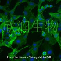
4. Mouse primary cultured renal vascular ECs and VSMCs were obtained from Newgainbio company, which were tested by Factor VIII and α-smooth muscle actin (α-SMA), the marker of ECs and VSMCs. RNeasy Mini Kit was used for RNA extraction, and the above protocols were repeated.
5. Porcine primary colon epithelial cells (Newgainbio company, Wuxi,China) were cultured in Dulbecco's Modified Eagle's Medium (Solarbio, Beijing, China) containing 10 % fetal bovine serum (BioInd, Kiryat shmona, Lsrael) at 37 ◦C and 5 % CO2 humidity.
Studying Osteocyte Function Using the Cell Lines MLO-Y4 and MLO-A5
We describe the culture and use of MLO-Y4 cells in studies of gene expression, response to fluid flow, and dendrite growth. We also describe how to use the MLO-A5 cells as a model of osteoblast to osteocyte �differentiation and how to study
三句话读懂一篇 CNS:生酮饮食有抗癌奇效?瘫痪患者福音,治疗 1 天即可恢复运动!
-edited powdery mildew resistance in wheat without growth penalties。 该研究通过叠加使用基因编辑技术,在敲除 MLO 感病基因的同时,删除了 TaMLO-B 附近的 304Kb 大片段 DNA,将抗病高产优异性状引入至小麦品种中,从此小麦不再惧怕白粉病! 图 1:来源 Nature 2. Cell Reports:发现肝星状细胞表达免疫检查点分子 PD-L1 肝内胆管癌是一种常见的恶性肝脏肿瘤,发病率仅次于肝癌。 2022
三句话读懂一篇 CNS:新疗法助力治疗女性不育;能负载活细胞的冷冻微针治疗技术;发现治疗心肌梗死新靶标,...
夏日好时光,本周学术君继续带你遨游学术之海,探寻科学的奥秘。 1. Nature Biomedical Engineering:首创冷冻微针治疗技术 细胞治疗,是一种能够将活细胞传递至体内发挥治疗效果的新型技术。 2021 年 5 月 3 日,香港城市大学徐臣杰教授研究团队在 Nature Biomedical Engineering 杂志上发表研究论文 Cryomicroneedles for Transdermal Cell Delivery。该工作首次创建能够负载活细胞的冷冻微针技术


