


- ¥1500
- ATCC、DSMZ、ECACC、RIKEN
- 江苏
- CL1354
- 2026年03月21日

企业认证
相关产品推荐更多 >


万千商家帮你免费找货
0 人在求购买到急需产品
- 详细信息
- 询价记录
- 文献和实验
- 技术资料
- 英文名:
A549
- 库存:
100万
- 供应商:
欣润生物
- 肿瘤类型:
是
- 细胞类型:
细胞系
- ATCC Number:
详见说明
- 品系:
人
- 组织来源:
肺
- 相关疾病:
肺癌
- 物种来源:
人源
- 免疫类型:
不详
- 细胞形态:
上皮型
- 是否是肿瘤细胞:
是
- 器官来源:
肺
- 运输方式:
新鲜或干冰
- 年限:
成年
- 生长状态:
贴壁生长
- 规格:
T25方瓶
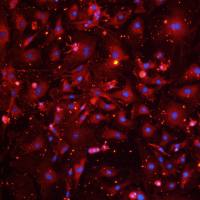
- 细胞名称:A549细胞(人肺癌细胞)
- 形态:上皮型,贴壁生长
- 含量:>1x10⁶ 个/瓶
- 污染:支原体、细菌、酵母和真菌检测为阴性
- 规格:T25瓶或者1mL冻存管包装
二、细胞接收后的处理:
1、贴壁细胞
- 收到T25方瓶细胞后,请检查是否漏液,如果漏液,请拍照片发给我们(冻存管细胞收到后直接37℃水浴复苏或直接放置于液氮中长期储存)。
- 请先在显微镜下确认细胞生长状态,去掉封口膜并将T25瓶置于37℃培养约2-3h。
- 弃去T25瓶中的培养基,换用新鲜的完全培养基。
- 如果细胞长满(90%以上)请及时进行细胞传代。
- 接到细胞次日,请检查细胞是否污染,若发现污染或疑似污染,请及时与我们取得联系。
2、悬浮细胞
- 收到细胞后,请检查是否漏液,如果漏液,请拍照片发给我们。
- 请先在显微镜下确认细胞生长状态,去掉封口膜并将15ml离心管置于37℃培养约2-3h。
- 1200rpm离心5min,弃去15ml离心管中的培养基,细胞沉淀用新鲜的完全培养基重悬并培养。
- 如果细胞长满(90%以上)请及时进行细胞传代。
- 接到细胞次日,请检查细胞是否污染,若发现污染或疑似污染,请及时与我们取得联系。
本公司的细胞培养操作规程,供参考
一、培养基及培养冻存条件准备:
- 准备F-12K培养基,90%;优质胎牛血清,10%。
- 培养条件: 气相:空气,95%;二氧化碳,5%。 温度:37℃,培养箱湿度为70%-80%。
- 冻存液:90%血清,10%DMSO,现用现配。液氮储存。
对于贴壁细胞,传代可参考以下方法:
- 弃去培养上清,用不含钙、镁离子的PBS润洗细胞1-2次。
- 加2ml消化液(0.25%Trypsin-0.53mM EDTA)于培养瓶中,置于37℃培养箱中消化2-3分钟,然后在显微镜下观察细胞消化情况,若细胞大部分变圆并脱落,迅速拿回操作台,轻敲几下培养瓶后加入3ml此细胞的培养基终止消化。
- 轻轻吹打后吸出,移入15ml离心管中,在1200RPM条件下离心5分钟,弃去上清液,加入1mL培养液后吹匀。
- 移入到事先准备好的含有5ml培养基的T-25培养瓶中或含有14ml培养基的T-75培养瓶中培养。
3)细胞冻存:待细胞生长状态良好时,可进行细胞冻存。贴壁细胞冻存时,先要消化处理并进行细胞计数。消化方法按照细胞传代方法的1-3步骤进行,最后的重悬液使用血清。悬浮细胞直接计数后离心,用血清重悬浮,加DMSO至最终浓度为10%。加入DMSO后迅速混匀,按每1ml的数量分配到冻存管中。本公司按每个冻存管细胞数目大于1X10⁶个细胞冻存。
注意事项:
1. 收到冻存管细胞后,若发现干冰已挥发干净、冻存管瓶盖脱落、破损及细胞有污染,请立即与我们联系。
2. 所有动物细胞均视为有潜在的生物危害性,必须在二级生物安全台内操作,并请注意防护,所有废液及接触过此细胞的器皿需要灭菌后方能丢弃。
3. 细胞用途:仅供科研使用。
发货方式:
复苏后发货:我们复苏细胞后发货,货期一周左右,免运费。(气温较好建议复苏后发货)
冻存发货(干冰运输):需额外增加干冰运费,选择干冰运输的我们发两管细胞,为了保证客户接种可靠性多发一管。(气温低于0℃须冻存发货)
细胞发货采取专业的运输包装,并选择最快捷的运输方式(顺丰速运或其他空运快递)
TGF-β1 exposure induces epithelial to mesenchymal transition both in CSCs and non-CSCs of the A549 cell line, leading to an increase of migration ability in the CD133+ A549 cell fraction
Metastasis is the leading cause of death by cancer. Non-small-cell lung cancer (NSCLC) represents nearly 85% of primary malignant lung tumours. Recent researches have demonstrated that epithelial-to-mesenchymal transition (EMT) plays a key role in the early process of metastasis of cancer cells. Transforming growth factor-β1 (TGF-β1) is the major inductor of EMT. The aim of this study is to investigate TGF-β1's effect on cancer stem cells (CSCs) identified as cells positive for CD133, side population (SP) and non-cancer stem cells (non-CSCs) identified as cells negative for CD133, and SP in the A549 cell line. We demonstrate that TGF-β1 induces EMT in both CSC and non-CSC A549 sublines, upregulating the expression of mesenchymal markers such as vimentin and Slug, and downregulating levels of epithelial markers such as e-cadherin and cytokeratins. CSC and non-CSC A549 sublines undergoing EMT show a strong migration and strong levels of MMP9 except for the CD133 cell fraction. OCT4 levels are strongly upregulated in all cell fractions except CD133 cells. On the contrary, wound size reveals that TGF-β1 enhances motility in wild-type A549 as well as CD133+ and SP+ cells. For CD133 and SP cells, TGF-β1 exposure does not change the motility. Finally, assessment of growth kinetics reveals major colony-forming efficiency in CD133+ A549 cells. In particular, SP+ and SP A549 cells show more efficiency to form colonies than untreated corresponding cells, while for CD133 cells no change in colony number was observable after TGF-β1 exposure. We conclude that it is possible to highlight different cell subpopulations with different grades of stemness. Each population seems to be involved in different biological mechanisms such as stemness maintenance, tumorigenicity, invasion and migration.
Characterization of the A549 cell line as a type II pulmonary epithelial cell model for drug metabolism.
Multiple cell types contribute to the pulmonary barrier including Type I and Type II alveolar epithelium. The objective of this research was to establish and characterize an in vitro model of Type II alveolar epithelium using the A549 human lung adenocarcinoma cell line. A549 cells form confluent monolayers with Type II characteristic morphology and tannic acid staining for typical lamellar bodies. A549 cells possess P450 IA1 and P450 IIB6 as determined by Western blots. Both CYPIA1 and CYPIIB6 P450 isozymes were determined to be functional with the fluorescent resorufin assay. Only the IA1 isozyme was observed to be inducible with selected polycyclic hydrocarbons. Uptake and transport experiments were carried out in cluster plates and in Snapwells. Cationized ferritin, a nonspecific absorbtive marker, was found to be taken up by the cells in a concentration-, time-, and temperature-dependent fashion. Lucifer yellow, a fluid-phase marker, was not internalized by the A549 cells. Transferrin, a representative receptor-mediated endocytic marker, was found to be taken up by the cells in a concentration-dependent and competitive fashion. Transport experiments involving fluorescein-transferrin also showed that A549 monolayers were polarized, with a greater amount of intracellular transferrin being transported out of the basolateral side of the cells. The experimental data agree favorably with literature for primary cultures of Type II pulmonary epithelial cells. These results indicated that the A549 cell line may be useful for the studying the metabolic and macromolecule processing contributions of alveolar Type II cells to mechanisms of drug delivery at the pulmonary epithelium. Copyright 1998 Academic Press.
风险提示:丁香通仅作为第三方平台,为商家信息发布提供平台空间。用户咨询产品时请注意保护个人信息及财产安全,合理判断,谨慎选购商品,商家和用户对交易行为负责。对于医疗器械类产品,请先查证核实企业经营资质和医疗器械产品注册证情况。
- 作者
- 内容
- 询问日期
 文献和实验
文献和实验Chicken intestinal epithelial cells were obtained from NEWGAINBIO company. Cells were cultured on 37℃, with 5% CO2, in the Ham’s F-12 Nutrient (DMEM/12) that contained the following supplementations: fetal bovine serum (5%), in-sulin (5 µg/mL), transferrin (5 µg/mL), selenium (5 ng/mL), epidermal growth factor (5 ng/mL) and penicillin-streptomycin (100–100 U/mL) for cell culturing (full DMEM/12). Experiments were performed with chicken intestinal epithelial cells and working solutions were prepared with plain DMEM/12 without supplementation. For the investigations, cells were seeded onto 96-well, 24-well or 6-well polystyrene cell culture plates.
Primary hVICs (passage 2) were cultured to 50–60% confluence and infected with pGMLV-SV40T-puro lentivirus (NewgainBio, Wuxi, China) at a multiplicity of infection of 80 supplemented with 5 µg/mL polybrene (Sigma-Aldrich, Buchs, Switzerland).
Tissue was cultured until cells became visible around the tissue, and when the fusion reached 90% (FIGURE 1A) §ask ¦lled with the prepared culturing medium was sent to the company for further immortalisation. Cell immortalisation was done for cell stability and longer-term use. Immortalised cells were cultured with 10% FBS and 1% PS in the DMEM medium. After the cells multiplied and merged, they were routinely passed and grown ( NEWGAINBIO Inc. Wuxi, Jiangsu, China) (FIGURE 1B-C).
Mouse primary cultured renal vascular ECs and VSMCs were obtained from Newgainbio company, which were tested by Factor VIII and α-smooth muscle actin (α-SMA), the marker of ECs and VSMCs. RNeasy Mini Kit was used for RNA extraction, and the above protocols were repeated.
Porcine primary colon epithelial cells (Newgainbio company, Wuxi,China) were cultured in Dulbecco's Modified Eagle's Medium (Solarbio, Beijing, China) containing 10 % fetal bovine serum (BioInd, Kiryat shmona, Lsrael) at 37 ◦C and 5 % CO2 humidity.
问: A549细胞到底是如何形态?我见过三角形,近乎MDCK细胞形态的,还有的呈长梭形。我感觉自己的细胞好像混进了MDCK细胞。大家帮忙诊断一下,谢谢。 我培养的A549细胞 答1:
我也在养A549细胞,也来讲讲自己的经验,希望大家共同学习。我们主要是看细胞的密度,大概有80-90%铺满培养瓶底就可以传代了,时间长短不一,大概有5-7天吧,一瓶一般传3-4瓶,中间一般会有一次换液的。培养基用的是1640+10%的小牛血清+200u/ml庆大霉素。传代时先倒掉就培养基,然后用PBS洗一两次,然后再用0.25%胰酶消化(100ml培养瓶盖住表面大概要两滴管左右),可镜经下观察细胞间已出现细胞胞质回缩变圆,缝隙变大(我的细胞大概要2min左右),即可倒去消化液,加入培养基,吹打
细胞也有身份证么?有啊,包括他的基因突变、表达、耐药,等等……要是不知道这些怎么选来做实验呢?
:也可以直接选,比如我们选一个A549细胞系:这里就会显示出A549细胞系的信息,包含有该细胞系的一系列数据库,比如药物敏感性的数据库,甲基化数据库,CRISPR敲除数据库,表达谱等等。首先会有一个总览,并且显示这些数据库的具体数据,数据都是可以下载的:右边显示的这个词云图,其实是和A549相关的主要突变驱动基因。点击信片数据的话,可以进入类似Array Express的界面,去下载数据:药物敏感,也是通过药物大规模筛选获得的结果:可以按照Z score来排序,找到A549最敏感的药物,以及靶点


