


- ¥1500
- ATCC、DSMZ、ECACC、RIKEN
- 江苏
- CL1076
- 2026年06月01日

企业认证
相关产品推荐更多 >


万千商家帮你免费找货
0 人在求购买到急需产品
- 详细信息
- 询价记录
- 文献和实验
- 技术资料
- 英文名:
Hacat
- 库存:
100万
- 供应商:
欣润生物
- 肿瘤类型:
否
- 细胞类型:
细胞系
- ATCC Number:
详见说明
- 品系:
人
- 组织来源:
角质形成层
- 相关疾病:
正常型
- 物种来源:
人源
- 免疫类型:
不详
- 细胞形态:
上皮型
- 是否是肿瘤细胞:
否
- 器官来源:
皮肤
- 运输方式:
新鲜或干冰
- 年限:
成年
- 生长状态:
贴壁生长
- 规格:
T25方瓶
一、细胞介绍
1)英文名称:Hacat细胞
(2)中文名称:人永生化角质形成细胞
(3)Hacat细胞:该细胞源自一位62岁人皮肤自发永生化角质形成层细胞,由于体外具有高度的增殖和分化能力,常被用于皮肤科学和分化研究中。各种慢些皮肤病研究中它们也是理解表皮稳态,病理生理学,T辅助细胞的作用机制的一种必要工具。角蛋白、角化细胞交联外膜蛋白、中间丝相关蛋白阳性。
(4)组织来源:人皮肤
(5)生长方式:贴壁生长
(6)细胞形态:上皮型
(7)规格:T25方瓶或1ml冻存管
(8)细胞数量:1×10^6
(9)Hacat细胞完全培养基:MEM+10%FBS+1%双抗
(10)培养条件:37℃,5%CO2
(11)运输方式:常温运输(T25方瓶)或干冰运输(冻存管)
(12)支原体检测:阴性
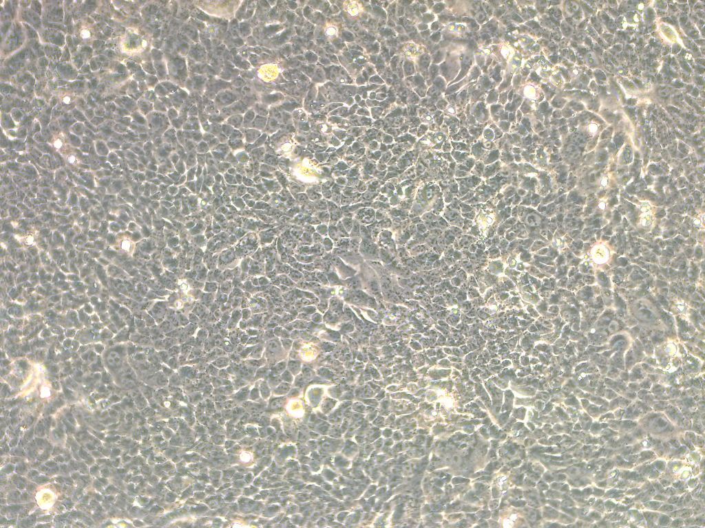
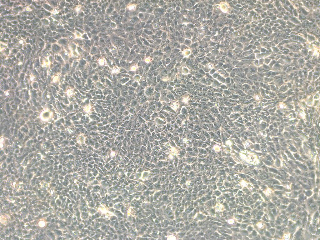
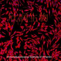
Hacat细胞白光图片


Cytotoxic Action of Juglone and Plumbagin: A Mechanistic Study Using HaCaT Keratinocytes
Juglone (5-hydroxy-1,4-naphthoquinone) and plumbagin (5-hydroxy-3-methyl-1,4-naphthoquinone) are yellow pigments found in black walnut (Juglans regia). Herbal preparations derived from black walnut have been used as hair dyes and skin colorants in addition to being applied topically for the treatment of acne, inflammatory diseases, ringworm, and fungal, bacterial, or viral infections. We have studied the cytotoxicity of these quinones to HaCaT keratinocytes. Exposure to juglone or plumbagin (1−20 μM) resulted in a concentration-dependent decrease in cell viability. The cytotoxicity of these quinones is due to two different mechanisms, namely, redox cycling and reaction with glutathione (GSH). Redox cycling results in the generation of the corresponding semiquinone radicals, which were detected by electron paramagnetic resonance. Incubation of keratinocytes with the quinones generated hydrogen peroxide (H2O2) and resulted in the oxidation of GSH to GS. Depletion of GSH by buthionine sulfoximine enhanced semiquinone radical production, increased H2O2 generation
风险提示:丁香通仅作为第三方平台,为商家信息发布提供平台空间。用户咨询产品时请注意保护个人信息及财产安全,合理判断,谨慎选购商品,商家和用户对交易行为负责。对于医疗器械类产品,请先查证核实企业经营资质和医疗器械产品注册证情况。
- 作者
- 内容
- 询问日期
 文献和实验
文献和实验Chicken intestinal epithelial cells were obtained from NEWGAINBIO company. Cells were cultured on 37℃, with 5% CO2, in the Ham’s F-12 Nutrient (DMEM/12) that contained the following supplementations: fetal bovine serum (5%), in-sulin (5 µg/mL), transferrin (5 µg/mL), selenium (5 ng/mL), epidermal growth factor (5 ng/mL) and penicillin-streptomycin (100–100 U/mL) for cell culturing (full DMEM/12). Experiments were performed with chicken intestinal epithelial cells and working solutions were prepared with plain DMEM/12 without supplementation. For the investigations, cells were seeded onto 96-well, 24-well or 6-well polystyrene cell culture plates.
Primary hVICs (passage 2) were cultured to 50–60% confluence and infected with pGMLV-SV40T-puro lentivirus (NewgainBio, Wuxi, China) at a multiplicity of infection of 80 supplemented with 5 µg/mL polybrene (Sigma-Aldrich, Buchs, Switzerland).
Tissue was cultured until cells became visible around the tissue, and when the fusion reached 90% (FIGURE 1A) §ask ¦lled with the prepared culturing medium was sent to the company for further immortalisation. Cell immortalisation was done for cell stability and longer-term use. Immortalised cells were cultured with 10% FBS and 1% PS in the DMEM medium. After the cells multiplied and merged, they were routinely passed and grown ( NEWGAINBIO Inc. Wuxi, Jiangsu, China) (FIGURE 1B-C).
Mouse primary cultured renal vascular ECs and VSMCs were obtained from Newgainbio company, which were tested by Factor VIII and α-smooth muscle actin (α-SMA), the marker of ECs and VSMCs. RNeasy Mini Kit was used for RNA extraction, and the above protocols were repeated.
Porcine primary colon epithelial cells (Newgainbio company, Wuxi,China) were cultured in Dulbecco's Modified Eagle's Medium (Solarbio, Beijing, China) containing 10 % fetal bovine serum (BioInd, Kiryat shmona, Lsrael) at 37 ◦C and 5 % CO2 humidity.
HaCaT 细胞中 PP6 后,通过 3D 培养,发现 PP6 的敲低能促进细胞表皮增生。 图 1 角质形成细胞中 PP6 的下调增加了皮肤的表皮厚度 接着,作者利用化合物库 Tsbiochem 筛选到化合物 L- 薄荷醇能显著提高 HaCaT 中 PP6的蛋白水平,但有趣的是,它并不影响细胞的活力和 PP6 的 mRNA 水平。作者利用体内实验探讨了 L- 薄荷醇对银屑病的治疗作用,发现与对照组相比,L- 薄荷醇减轻了小鼠耳朵的炎症表型(红斑、厚度和脱屑),同时减少了损伤皮肤的表皮厚度、细胞
利用 Millicell 培养小室培养皮肤及肺类器官的实验方案
气液界面培养(ALI)3D培养类器官实验方案- 皮肤类器官和肺类器官 1. 生成3D 皮肤类器官作为人体表皮的体外模型 皮肤是人体最大的器官,具有多种功能,包括保护、吸收物质和调节体温。皮肤具有复杂的分层结构,由三个主要层组成:1) 表皮紧密排列的细胞形成了防止物质入侵和水分流失的屏障,并包括一个称为角质层的外层,由重叠的、无活的角质细胞组成,可防止例如紫外线辐射和病原体的威胁;2)真皮,它支持着表皮,包含神经末梢、汗腺、皮脂腺、毛囊和血管。真皮的基底层是由角质形成细胞不断进行有丝分裂以补充
形成层和初生木质部三部分组成。注意韧皮部和木质部的排列方式与根有何不同?② 髓射线为各维管束之间的薄壁组织,是连接皮层与髓的薄壁组织。③ 髓维管束内方,茎的中央部分,由大量圆形的薄壁细胞组成。2.单子叶植物茎的初生构造 单子叶植物大多数只有初生结构,没有次生结构。取玉米和小麦茎横切制片(图6-4,图6-5,图6-6),在显微镜下观察 (1)表皮为茎最外面的一层细胞,其外被有角质层。 (2)下表层为表皮下方的几层小型细胞或厚壁细胞。 (3)基本组织为下皮层以内的所有薄壁组织,细胞较大


