


- ¥780 - 2200
- LMAI Bio
- LM-6983R
- 进口/国产
- 2026年05月19日
- WB=1:500-2000 ELISA=1:500-1000 IHC-P=1:400-800 IHC-F=1:400-800 Flow-Cyt=1ug/test IF=1:100-500 (石蜡切片需做抗原修复)
- Rabbit
- Human, Mouse, Rat, Chicken, Dog, Pig, Cow, Sheep,

企业认证
相关产品推荐更多 >


万千商家帮你免费找货
0 人在求购买到急需产品
- 详细信息
- 文献和实验
- 技术资料
- 供应商:
上海联迈生物工程有限公司
- 库存:
大量
- 目录编号:
LM-6983R
- 克隆性:
多克隆
- 抗原来源:
Rabbit
- 保质期:
1年
- 抗体英文名:
Wilms Tumor Protein
- 抗体名:
肾母细胞瘤蛋白抗体
- 宿主:
Rabbit
- 适应物种:
Human, Mouse, Rat, Chicken, Dog, Pig, Cow, Sheep,
- 免疫原:
KLH conjugated synthetic peptide derived from human Wilms Tumor Protein:301-400/449
- 亚型:
IgG
- 形态:
Lyophilized or Liquid
- 应用范围:
WB=1:500-2000 ELISA=1:500-1000 IHC-P=1:400-800 IHC-F=1:400-800 Flow-Cyt=1ug/test IF=1:100-500 (石蜡切片需做抗原修复)
- 浓度:
1mg/ml
- 保存条件:
Store at -20 °C
- 规格:
50ul 100ul 200ul
| 英文名称 | Wilms Tumor Protein |
| 中文名称 | 肾母细胞瘤蛋白抗体 |
| 别 名 | WIT 2; WT 1; AWT1; FWT1; GUD; NPHS4; WAGR; Wilms tumor 1; Wilms Tumor; Wilms tumor protein; Wilms' tumor gene; Wilms' tumor protein; WIT2; WT; WT1; WT-1; WT1_HUMAN; WT33. |
 |
Specific References (1) | bs-6983R has been referenced in 1 publications.
[IF=2.33] Xiao, Tangli, et al. "Rapamycin promotes podocyte autophagy and ameliorates renal injury in diabetic mice." Molecular and Cellular Biochemistry (2014): 1-10. IHC-P ; Mouse.
PubMed:24850187
|
| 规格价格 | 50ul/780元 购买 100ul/1380元 购买 200ul/2200元 购买 大包装/询价 |
| 说 明 书 | 50ul 100ul 200ul |
| 研究领域 | 肿瘤 细胞生物 免疫学 发育生物学 肿瘤细胞生物标志物 表观遗传学 |
| 抗体来源 | Rabbit |
| 克隆类型 | Polyclonal |
| 交叉反应 | Human, Mouse, Rat, Chicken, Dog, Pig, Cow, Sheep, |
| 产品应用 | WB=1:500-2000 ELISA=1:500-1000 IHC-P=1:400-800 IHC-F=1:400-800 Flow-Cyt=1ug/test IF=1:100-500 (石蜡切片需做抗原修复) not yet tested in other applications. optimal dilutions/concentrations should be determined by the end user. |
| 分 子 量 | 55kDa |
| 细胞定位 | 细胞核 细胞浆 |
| 性 状 | Lyophilized or Liquid |
| 浓 度 | 1mg/ml |
| 免 疫 原 | KLH conjugated synthetic peptide derived from human Wilms Tumor Protein:301-400/449 |
| 亚 型 | IgG |
| 纯化方法 | affinity purified by Protein A |
| 储 存 液 | 0.01M TBS(pH7.4) with 1% BSA, 0.03% Proclin300 and 50% Glycerol. |
| 保存条件 | Store at -20 °C for one year. Avoid repeated freeze/thaw cycles. The lyophilized antibody is stable at room temperature for at least one month and for greater than a year when kept at -20°C. When reconstituted in sterile pH 7.4 0.01M PBS or diluent of antibody the antibody is stable for at least two weeks at 2-4 °C. |
| PubMed | PubMed |
| 产品介绍 | background: Transcription factor that plays an important role in cellular development and cell survival. Regulates the expression of numerous target genes, including EPO. Plays an essential role for development of the urogenital system. Recognizes and binds to the DNA sequence 5'-CGCCCCCGC-3'. It has a tumor suppressor as well as an oncogenic role in tumor formation. Function may be isoform-specific: isoforms lacking the KTS motif may act as transcription factors. Isoforms containing the KTS motif may bind mRNA and play a role in mRNA metabolism or splicing. Isoform 1 has lower affinity for DNA, and can bind RNA. Function: Transcription factor that plays an important role in cellular development and cell survival. Regulates the expression of numerous target genes, including EPO. Plays an essential role for development of the urogenital system. Recognizes and binds to the DNA sequence 5'-CGCCCCCGC-3'. It has a tumor suppressor as well as an oncogenic role in tumor formation. Function may be isoform-specific: isoforms lacking the KTS motif may act as transcription factors. Isoforms containing the KTS motif may bind mRNA and play a role in mRNA metabolism or splicing. Isoform 1 has lower affinity for DNA, and can bind RNA. Subunit: Homodimer. Interacts with WTIP. Interacts with actively translating polysomes. Detected in nuclear ribonucleoprotein (mRNP) particles. Interacts with HNRNPU via the zinc-finger region. Interacts with U2AF2. Interacts with CITED2. Interacts with ZNF224 via the zinc-finger region. Interacts with WTAP and SRY. Interacts with FAM123B/WTX. Interacts with RBM4. Subcellular Location: Nucleus. Nucleus, nucleolus. Cytoplasm. Note=Shuttles between nucleus and cytoplasm. Isoform 1: Nucleus speckle. Isoform 4: Nucleus, nucleoplasm. Tissue Specificity: Expressed in the kidney and a subset of hematopoietic cells. DISEASE: Defects in WT1 are the cause of Frasier syndrome (FS) [MIM:136680]. FS is characterized by a slowly progressing nephropathy leading to renal failure in adolescence or early adulthood, male pseudohermaphroditism, and no Wilms tumor. As for histological findings of the kidneys, focal glomerular sclerosis is often observed. There is phenotypic overlap with Denys-Drash syndrome. Inheritance is autosomal dominant. Defects in WT1 are the cause of Wilms tumor 1 (WT1) [MIM:194070]. WT is an embryonal malignancy of the kidney that affects approximately 1 in 10'000 infants and young children. It occurs both in sporadic and hereditary forms. Defects in WT1 are the cause of Denys-Drash syndrome (DDS) [MIM:194080]. DDS is a typical nephropathy characterized by diffuse mesangial sclerosis, genital abnormalities, and/or Wilms tumor. There is phenotypic overlap with WAGR syndrome and Frasier syndrome. Inheritance is autosomal dominant, but most cases are sporadic. Similarity: Belongs to the EGR C2H2-type zinc-finger protein family. Contains 4 C2H2-type zinc fingers. SWISS: P19544 Gene ID: 7490 Database links: Entrez Gene: 7490 Human Entrez Gene: 22431 Mouse Entrez Gene: 24883 Rat Omim: 607102 Human SwissProt: P19544 Human SwissProt: P22561 Mouse SwissProt: P49952 Rat Unigene: 591980 Human Unigene: 389339 Mouse Unigene: 92531 Rat Important Note: This product as supplied is intended for research use only, not for use in human, therapeutic or diagnostic applications. |
| 产品图片 |
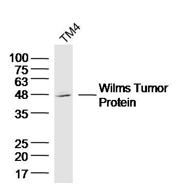
Sample: TM4 Cell (Mouse) Lysate at 40 ug
Primary: Anti-Wilms Tumor Protein (bs-6983R) at 1/300 dilution Secondary: IRDye800CW Goat Anti-Rabbit IgG at 1/20000 dilution Predicted band size: 55 kD Observed band size: 48 kD 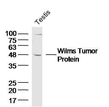
Sample: Testis (Mouse) Lysate at 40 ug
Primary: Anti-Wilms Tumor Protein (bs-6983R) at 1/300 dilution Secondary: IRDye800CW Goat Anti-Rabbit IgG at 1/20000 dilution Predicted band size: 55 kD Observed band size: 48 kD 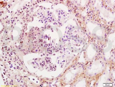
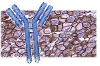
Tissue/cell: human kidney tissue; 4% Paraformaldehyde-fixed and paraffin-embedded;
Antigen retrieval: citrate buffer ( 0.01M, pH 6.0 ), Boiling bathing for 15min; Block endogenous peroxidase by 3% Hydrogen peroxide for 30min; Blocking buffer (normal goat serum,C-0005) at 37℃ for 20 min; Incubation: Anti-WT-1/Wilms Tumor Protein Polyclonal Antibody, Unconjugated(bs-6983R) 1:200, overnight at 4°C, followed by conjugation to the secondary antibody(SP-0023) and DAB(C-0010) staining 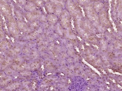
Paraformaldehyde-fixed, paraffin embedded (Mouse kidney); Antigen retrieval by boiling in sodium citrate buffer (pH6.0) for 15min; Block endogenous peroxidase by 3% hydrogen peroxide for 20 minutes; Blocking buffer (normal goat serum) at 37°C for 30min; Antibody incubation with (Wilms Tumor Protein) Polyclonal Antibody, Unconjugated (bs-6983R) at 1:400 overnight at 4°C, followed by operating according to SP Kit(Rabbit) (sp-0023) instructionsand DAB staining.
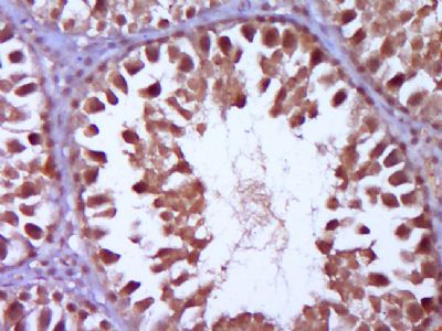
Paraformaldehyde-fixed, paraffin embedded (Rat testis); Antigen retrieval by boiling in sodium citrate buffer (pH6.0) for 15min; Block endogenous peroxidase by 3% hydrogen peroxide for 20 minutes; Blocking buffer (normal goat serum) at 37°C for 30min; Antibody incubation with (Wilms Tumor Protein) Polyclonal Antibody, Unconjugated (bs-6983R) at 1:400 overnight at 4°C, followed by operating according to SP Kit(Rabbit) (sp-0023) instructionsand DAB staining.
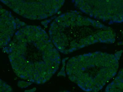
Paraformaldehyde-fixed, paraffin embedded (Mouse testis); Antigen retrieval by boiling in sodium citrate buffer (pH6.0) for 15min; Block endogenous peroxidase by 3% hydrogen peroxide for 20 minutes; Blocking buffer (normal goat serum) at 37°C for 30min; Antibody incubation with (Wilms Tumor Protein) Polyclonal Antibody, Unconjugated (bs-6983R) at 1:400 overnight at 4°C, followed by a conjugated Goat Anti-Rabbit IgG antibody (bs-0295G-FITC) for 90 minutes, and DAPI for nuclei staining.
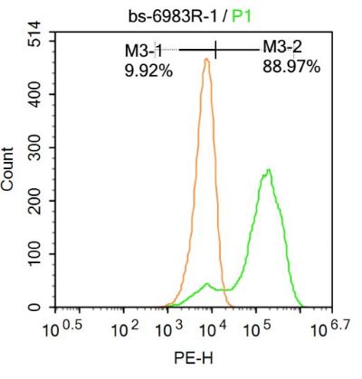
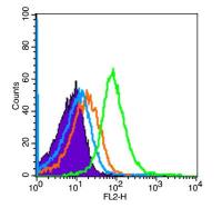
Blank control:Molt-4.
Primary Antibody (green line): Rabbit Anti-Wilms Tumor Protein antibody (bs-6983R) Dilution: 1μg /10^6 cells; Isotype Control Antibody (orange line): Rabbit IgG . Secondary Antibody : Goat anti-rabbit IgG-AF647 Dilution: 1μg /test. Protocol The cells were fixed with 4% PFA (10min at room temperature)and then permeabilized with 90% ice-cold methanol for 20 min at-20℃. The cells were then incubated in 5%BSA to block non-specific protein-protein interactions for 30 min at at room temperature .Cells stained with Primary Antibody for 30 min at room temperature. The secondary antibody used for 40 min at room temperature. Acquisition of 20,000 events was performed. |
风险提示:丁香通仅作为第三方平台,为商家信息发布提供平台空间。用户咨询产品时请注意保护个人信息及财产安全,合理判断,谨慎选购商品,商家和用户对交易行为负责。对于医疗器械类产品,请先查证核实企业经营资质和医疗器械产品注册证情况。
 文献和实验
文献和实验Comparative Genomic Hybridization of Wilms tumor
Cytogenetic analysis of solid tumors including Wilms’ tumor is challenging due to poor chromosome morphology, complexity of abnormalities, and to the possibility of stromal cell overgrowth in tissue culture. Molecular cytogenetic techniques
Wilms Tumor as a Model for Cancer Biology
Wilms’ tumor of the kidney (WT) is the most common solid tumor of childhood, and it was first described in detail by Max Wilms’ in 1899. WT is a paradigm of childhood cancer, because it has served as a model from four distinct perspectives
Mutational Analysis of the Wilms' Tumor (WTI) Gene
Mutations of the Wilms’ tumor (WT1) gene have been shown to underlie a proportion of cases of Wilms’ tumor, an embryonal kidney cancer occurring mainly in childhood. The WTl gene comprtses ten exons spanning approx 50 kb of genomrc DNA
 技术资料
技术资料暂无技术资料 索取技术资料

