


- ¥780 - 2200
- LMAI Bio
- LM-2556R
- 进口/国产
- 2026年03月26日
- WB=1:500-2000 ELISA=1:500-1000 IHC-F=1:400-800 Flow-Cyt=3ug/Test IF=1:100-500 (石蜡切片需做抗原修复)
- Rabbit
- Human, Mouse, Rat, Cow, Horse, Rabbit,

企业认证
相关产品推荐更多 >


万千商家帮你免费找货
0 人在求购买到急需产品
- 详细信息
- 文献和实验
- 技术资料
- 供应商:
上海联迈生物工程有限公司
- 库存:
大量
- 目录编号:
LM-2556R
- 克隆性:
多克隆
- 抗原来源:
Rabbit
- 保质期:
1年
- 抗体英文名:
LAMP3
- 抗体名:
溶酶体相关膜蛋白3抗体
- 宿主:
Rabbit
- 适应物种:
Human, Mouse, Rat, Cow, Horse, Rabbit,
- 免疫原:
KLH conjugated synthetic peptide derived from human LAMP-3:301-416/416
- 亚型:
IgG
- 形态:
Lyophilized or Liquid
- 应用范围:
WB=1:500-2000 ELISA=1:500-1000 IHC-F=1:400-800 Flow-Cyt=3ug/Test IF=1:100-500 (石蜡切片需做抗原修复)
- 浓度:
1mg/ml
- 保存条件:
Store at -20 °C
- 规格:
50ul 100ul 200ul
| 英文名称 | LAMP3 |
| 中文名称 | 溶酶体相关膜蛋白3抗体 |
| 别 名 | DC-lysosome-associated membrane glycoprotein; CD208; CD208 antigen; DC LAMP; DCLAMP; DC-LAMP; LAMP; LAMP 3; LAMP-3; LAMP3; Lysosomal associated membrane protein 3; Protein TSC403; TSC403; DC lysosome associated membrane glycoprotein; LAMP3_HUMAN; Lysosomal-associated membrane protein 3; Lysosome-associated membrane glycoprotein 3. |
| 规格价格 | 50ul/780元 购买 100ul/1380元 购买 200ul/2200元 购买 大包装/询价 |
| 说 明 书 | 50ul 100ul 200ul |
| 研究领域 | 肿瘤 干细胞 |
| 抗体来源 | Rabbit |
| 克隆类型 | Polyclonal |
| 交叉反应 | Human, Mouse, Rat, Cow, Horse, Rabbit, |
| 产品应用 | WB=1:500-2000 ELISA=1:500-1000 IHC-F=1:400-800 Flow-Cyt=3ug/Test IF=1:100-500 (石蜡切片需做抗原修复) not yet tested in other applications. optimal dilutions/concentrations should be determined by the end user. |
| 分 子 量 | 44kDa |
| 细胞定位 | 细胞浆 |
| 性 状 | Lyophilized or Liquid |
| 浓 度 | 1mg/ml |
| 免 疫 原 | KLH conjugated synthetic peptide derived from human LAMP-3:301-416/416 |
| 亚 型 | IgG |
| 纯化方法 | affinity purified by Protein A |
| 储 存 液 | 0.01M TBS(pH7.4) with 1% BSA, 0.03% Proclin300 and 50% Glycerol. |
| 保存条件 | Store at -20 °C for one year. Avoid repeated freeze/thaw cycles. The lyophilized antibody is stable at room temperature for at least one month and for greater than a year when kept at -20°C. When reconstituted in sterile pH 7.4 0.01M PBS or diluent of antibody the antibody is stable for at least two weeks at 2-4 °C. |
| PubMed | PubMed |
| 产品介绍 | background: LAMP3 might change the lysosome function after the transfer of peptide MHC class II molecules to the surface of dendritic cells. It's over expression is associated with an enhanced metastatic potential and may be a prognostic factor for cervical cancer. Expressed in lymphoid organs and dendritic cells and lung. Up regulated in carcinomas of the esophagus, colon, rectum, ureter, stomach, breast, fallopian tube, thyroid and parotid tissues. Function: Might change the lysosome function after the transfer of peptide-MHC class II molecules to the surface of dendritic cells. Subcellular Location: Lysosome membrane. Tissue Specificity: Expressed in lymphoid organs and dendritic cells. Expressed in lung. Up-regulated in carcinomas of the esophagus, colon, rectum, ureter, stomach, breast, fallopian tube, thyroid and parotid tissues. Similarity: Belongs to the LAMP family. SWISS: Q9UQV4 Gene ID: 27074 Database links: Entrez Gene: 27074 Human Omim: 605883 Human SwissProt: Q9UQV4 Human Unigene: 518448 Human Important Note: This product as supplied is intended for research use only, not for use in human, therapeutic or diagnostic applications. |
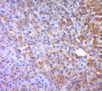
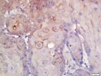
| 产品图片 | 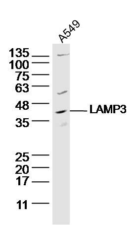 Sample: A549 Cell (Human) Lysate at 40 ug Primary: Anti- LAMP3 (bs-2556R) at 1/300 dilution Secondary: IRDye800CW Goat Anti-Rabbit IgG at 1/20000 dilution Predicted band size: 44 kD Observed band size: 44 kD 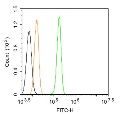 Blank control (Black line): A431 (Black). Primary Antibody (green line): Rabbit Anti-LAMP3 antibody (bs-2556R) Dilution: 1μg /10^6 cells; Isotype Control Antibody (orange line): Rabbit IgG . Dilution: 3μg /test. Protocol The cells were fixed with 4% PFA (10min at room temperature)and then permeabilized with 90% ice-cold methanol for 20 min at room temperature. The cells were then incubated in 5%BSA to block non-specific protein-protein interactions for 30 min at room temperature .Cells stained with Primary Antibody for 30 min at room temperature. Acquisition of 20,000 events was performed. |
风险提示:丁香通仅作为第三方平台,为商家信息发布提供平台空间。用户咨询产品时请注意保护个人信息及财产安全,合理判断,谨慎选购商品,商家和用户对交易行为负责。对于医疗器械类产品,请先查证核实企业经营资质和医疗器械产品注册证情况。
 文献和实验
文献和实验,内含胞浆成分,如线粒体、内质网、核糖体等。 自噬溶酶体(AV2)的特征为:单层膜,胞浆成分已降解。(autophagic vacuole,AV) 2. 在荧光显微镜下采用 GFP-LC3 融合蛋白来示踪自噬形成 由于电镜耗时长,不利于监测(Monitoring)自噬形成,人们利用 LC3 在自噬形成过程中发生聚集的现象开发出了此技术。无自噬时,GFP-LC3 融合蛋白弥散在胞浆中;自噬形成时,GFP-LC3 融合蛋白转位至自噬体膜,在荧光显微镜下形成多个明亮的绿色荧光斑点,一个斑
相关专题 Western是利用抗体检测蛋白常用的方法,本手册旨在表明Western-blot的技术原理、操作步骤及常见问题分析,可采用以下步骤进行Western-blot实验: Western-blot技术原理: Western Blot技术是通过聚丙烯酰胺凝胶电泳后,经PAGE分离的蛋白质样品,转移到固相载体(例如硝酸纤维素薄膜)上,固相载体以非共价键形式吸附蛋白质,且能保持电泳分离的多肽类型及其生物学活性不变。以固相载体
(RabS)作用下与溶酶体融合形成自噬溶酶体的过程,有些溶酶体相关蛋白也参与了其中,包括 LAMP1、LAMP2、UVRAG (紫外线抵抗相关肿瘤抑制基因)。而在降解阶段自噬体蛋白也发挥了重要作用,如 Atg22、Atg12、Atg15、Atg1、Atg13 等。细胞自噬的调节是一个多步骤的极其复杂的过程,目前机制尚未完全掌握,已知它主要是通过mTOR 信号通路、Beclin-1 信号通路、P53 信号通路三条途径来进行调控的。 1) mTOR 信号通路:mTOR 是是一个丝/苏氨酸
 技术资料
技术资料暂无技术资料 索取技术资料

