

- ¥1380 - 2200
- LMAI Bio
- LM-14629R
- 进口/国产
- 2026年09月28日
- WB=1:500-2000 ELISA=1:500-1000 IHC-P=1:400-800 IHC-F=1:400-800 ICC=1:100-500 IF=1:100-500 (石蜡切片需做抗原修复)
- Rabbit
- Human, Mouse, Rat, Chicken, Rabbit,

企业认证
相关产品推荐更多 >


万千商家帮你免费找货
0 人在求购买到急需产品
相关阅读
AJE 润色服务限时 8 折起,权威机构信任 15+ 年Cytoscape 教程来了!快速实现顶刊同款通路网络图五大应用案例:Mustang Q 膜层析应用全解析1 个小工具,一次性搞定流程图、质粒图谱和信号通路图- 详细信息
- 文献和实验
- 技术资料
- 供应商:
上海联迈生物工程有限公司
- 库存:
大量
- 目录编号:
LM-14629R
- 克隆性:
多克隆
- 抗原来源:
Rabbit
- 保质期:
1年
- 抗体英文名:
ERp57
- 抗体名:
内质网蛋白57抗体
- 宿主:
Rabbit
- 适应物种:
Human, Mouse, Rat, Chicken, Rabbit,
- 免疫原:
KLH conjugated synthetic peptide derived from human ERp57:351-450/505
- 亚型:
IgG
- 形态:
Lyophilized or Liquid
- 应用范围:
WB=1:500-2000 ELISA=1:500-1000 IHC-P=1:400-800 IHC-F=1:400-800 ICC=1:100-500 IF=1:100-500 (石蜡切片需做抗原修复)
- 浓度:
1mg/ml
- 保存条件:
Store at -20 °C
- 规格:
100ul 200ul
| 英文名称 | ERp57 |
| 中文名称 | 内质网蛋白57抗体 |
| 别 名 | ERp57/ERp60; 58 kDa glucose regulated protein; 58 kDa glucose-regulated protein; 58 kDa microsomal protein; Disulfide isomerase ER 60; Disulfide isomerase ER-60; Endoplasmic reticulum resident protein 57; Endoplasmic reticulum resident protein 60; ER p57; ER protein 57; ER protein 60; ERp 57; ERp57; ERp60; ERp61; Glucose Regulated Protein 58 Kd; GRP 57; GRP 58; GRP57; GRP58; HsT17083; P58; PDIA 3; PDIA3; PDIA3_HUMAN; Phospholipase C alpha; PI PLC; Protein disulfide isomerase A3; Protein disulfide isomerase family A member 3; Protein disulfide-isomerase A3. |
| 规格价格 | 100ul/1380元 购买 200ul/2200元 购买 大包装/询价 |
| 说 明 书 | 100ul 200ul |
| 研究领域 | 细胞生物 转运蛋白 糖蛋白 |
| 抗体来源 | Rabbit |
| 克隆类型 | Polyclonal |
| 交叉反应 | Human, Mouse, Rat, Chicken, Rabbit, |
| 产品应用 | WB=1:500-2000 ELISA=1:500-1000 IHC-P=1:400-800 IHC-F=1:400-800 ICC=1:100-500 IF=1:100-500 (石蜡切片需做抗原修复) not yet tested in other applications. optimal dilutions/concentrations should be determined by the end user. |
| 分 子 量 | 54kDa |
| 细胞定位 | 细胞浆 |
| 性 状 | Lyophilized or Liquid |
| 浓 度 | 1mg/ml |
| 免 疫 原 | KLH conjugated synthetic peptide derived from human ERp57:351-450/505 |
| 亚 型 | IgG |
| 纯化方法 | affinity purified by Protein A |
| 储 存 液 | 0.01M TBS(pH7.4) with 1% BSA, 0.03% Proclin300 and 50% Glycerol. |
| 保存条件 | Store at -20 °C for one year. Avoid repeated freeze/thaw cycles. The lyophilized antibody is stable at room temperature for at least one month and for greater than a year when kept at -20°C. When reconstituted in sterile pH 7.4 0.01M PBS or diluent of antibody the antibody is stable for at least two weeks at 2-4 °C. |
| PubMed | PubMed |
| 产品介绍 | background: This gene encodes a protein of the endoplasmic reticulum that interacts with lectin chaperones calreticulin and calnexin to modulate folding of newly synthesized glycoproteins. The protein was once thought to be a phospholipase; however, it has been demonstrated that the protein actually has protein disulfide isomerase activity. It is thought that complexes of lectins and this protein mediate protein folding by promoting formation of disulfide bonds in their glycoprotein substrates. [provided by RefSeq, Jul 2008] Subcellular Location: Endoplasmic reticulum lumen. Melanosome. Identified by mass spectrometry in melanosome fractions from stage I to stage IV. Tissue Specificity: Detected in the flagellum and head region of spermatozoa (at protein level). Similarity: Belongs to the protein disulfide isomerase family. Contains 2 thioredoxin domains. SWISS: P30101 Gene ID: 2923 Database links: Entrez Gene: 2923 Human Entrez Gene: 14827 Mouse Entrez Gene: 100328685 Rabbit Entrez Gene: 29468 Rat Omim: 602046 Human SwissProt: P30101 Human SwissProt: P27773 Mouse SwissProt: P11598 Rat Unigene: 591095 Human Unigene: 263177 Mouse Unigene: 11527 Rat Important Note: This product as supplied is intended for research use only, not for use in human, therapeutic or diagnostic applications. |
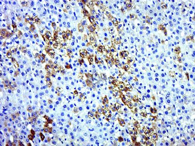
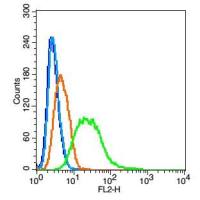
| 产品图片 | 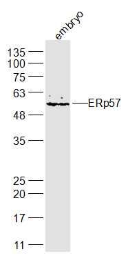 Sample: embryo (Mouse) Lysate at 40 ug Primary: Anti-ERp57 (bs-14629R) at 1/300 dilution Secondary: IRDye800CW Goat Anti-Rabbit IgG at 1/20000 dilution Predicted band size: 54 kD Observed band size: 54 kD 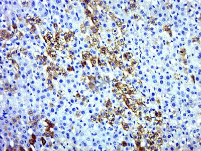 Paraformaldehyde-fixed, paraffin embedded (Rat liver); Antigen retrieval by boiling in sodium citrate buffer (pH6.0) for 15min; Block endogenous peroxidase by 3% hydrogen peroxide for 20 minutes; Blocking buffer (normal goat serum) at 37°C for 30min; Antibody incubation with (ERp57) Polyclonal Antibody, Unconjugated (bs-14629R) at 1:500 overnight at 4°C, followed by a conjugated secondary (sp-0023) for 20 minutes and DAB staining. |
风险提示:丁香通仅作为第三方平台,为商家信息发布提供平台空间。用户咨询产品时请注意保护个人信息及财产安全,合理判断,谨慎选购商品,商家和用户对交易行为负责。对于医疗器械类产品,请先查证核实企业经营资质和医疗器械产品注册证情况。
 文献和实验
文献和实验更加明显,RA 持续时间更长,但其起病较缓慢,没有病情的波动和复发情况,也没有关节外皮肤及眼部表现的改变。 图为集萃药康 Male DBA/1J 诱导模型 3. 胶原抗体诱导的关节炎(CAIA) CAIA 属于 CIA 模型的扩展,是抗体诱导型关节炎模型中最常见的模型之一。与 CIA 依赖的 MHC 单倍型易感品系相比,CAIA 可在大多数小鼠上诱导关节炎,如 DBA/1、BALB/c、C57BL/6、C57BL/10 等小鼠[8]。 造模机制:通过靶向 Ⅱ 型胶原蛋白的各种表位的抗体混合
Science 背靠背!施一公、黄志伟团队同日揭开 B 细胞受体复合物的神秘面纱
载体上,同时使用共表达方式,通过表达内质网蛋白 pERp1,pERp1 可以促进 IgM 二硫键的形成,二硫键有助于 IgM-BCR 正确折叠和结构稳定。 在完成蛋白的表达和纯化后,在溶液中加入抗体偶联药物 Polatuzumab vedotin 的 Fab 片段,最后通过冷冻电镜解析了第一个人源 IgM 同种型 B 细胞受体复合物 3.3Å 的高分辨率结构。 这篇文章所获得的结构,包含了复合物几乎胞外结构域以及所有跨膜区域, 包含一个 mIgM 和一个 Igα/Igβ 异源二聚体复合物。除此之外
实验室中时常听到这样的困惑,「明明通过免疫荧光(IF)能看到目的蛋白表达在细胞核膜上的表达,但为什么不论用总蛋白还是核蛋白进行 WB 检测,都无法检测到目的蛋白?」 类似这样的问题,你是否也深有感触? 实际操作过程中,是否遇到过免疫荧光有结果而蛋白印迹却没条带的情况?什么原因导致了这种情况?又该如何处理? 一、IF & WB 免疫荧光(IF)和免疫印迹(WB)都是利用抗原和抗体的特异性结合对目的蛋白质进行检测。IF 采用抗体和荧光检测固定细胞或组织中目的蛋白
 技术资料
技术资料暂无技术资料 索取技术资料

