


- ¥580 - 4680
- Bioss

- bs-0061R
- 2025年10月01日
- 产品信息以Bioss网站为准

企业认证

研选同类产品更多 >


万千商家帮你免费找货
0 人在求购买到急需产品
- 详细信息
- 用户评价
- 文献和实验
- 技术资料
- 应用范围:
产品信息以Bioss网站为准
- 规格:
100ul/500ul/200ul/1000ul
| 规格: | 100ul | 产品价格: | ¥580.0 |
|---|---|---|---|
| 规格: | 500ul | 产品价格: | ¥2280.0 |
| 规格: | 200ul | 产品价格: | ¥980.0 |
| 规格: | 1000ul | 产品价格: | ¥4680.0 |
| 产品编号 | bs-0061R |
| 英文名称 | beta-Actin Rabbit pAb, Loading Control |
| 中文名称 | β-肌动蛋白/β-Actin(内参)抗体 |
| 英文别名 | ACTB_HUMAN; Actin, cytoplasmic 1; ACTB; EC:3.6.4.- ; Beta actin; Actin, cytoplasmic 1, N-terminally processed; actin beta; BNS; CSMH; DDS1; THC8; BKRNS; BRWS1; PS1TP5BP1; Actin cytoplasmic 1; β actin; β-actin; |
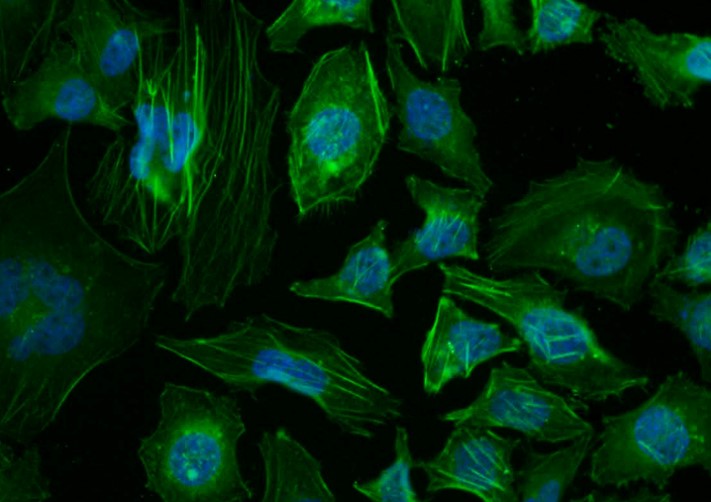
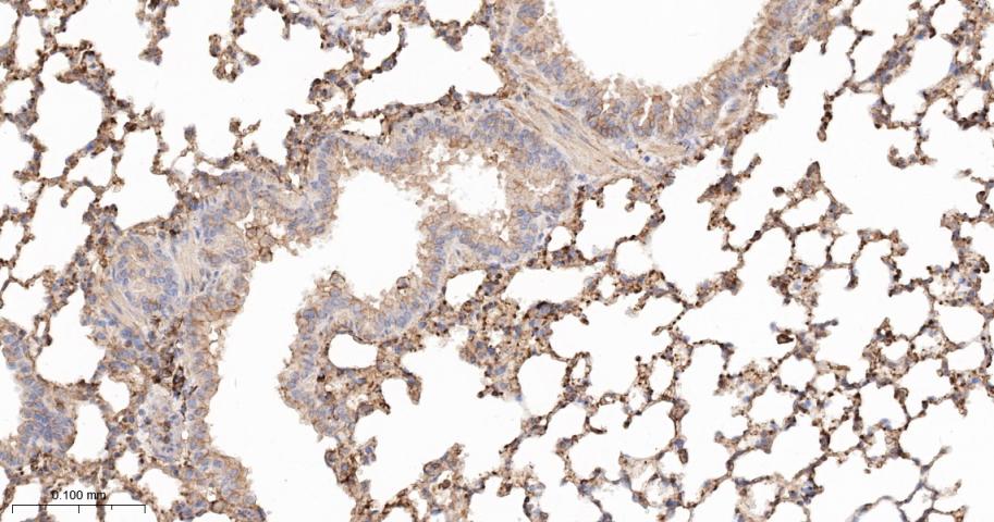
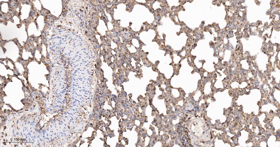
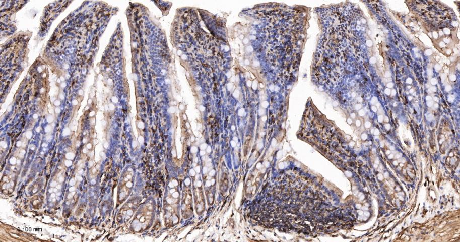
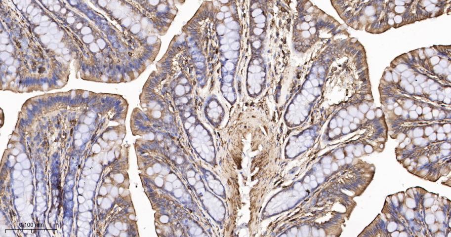
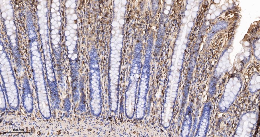
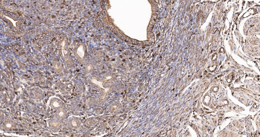
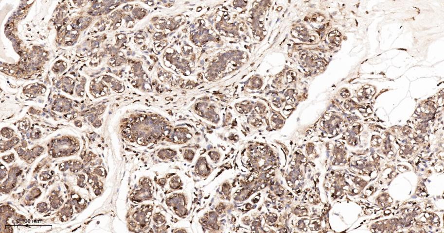
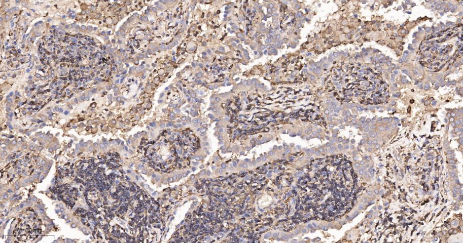
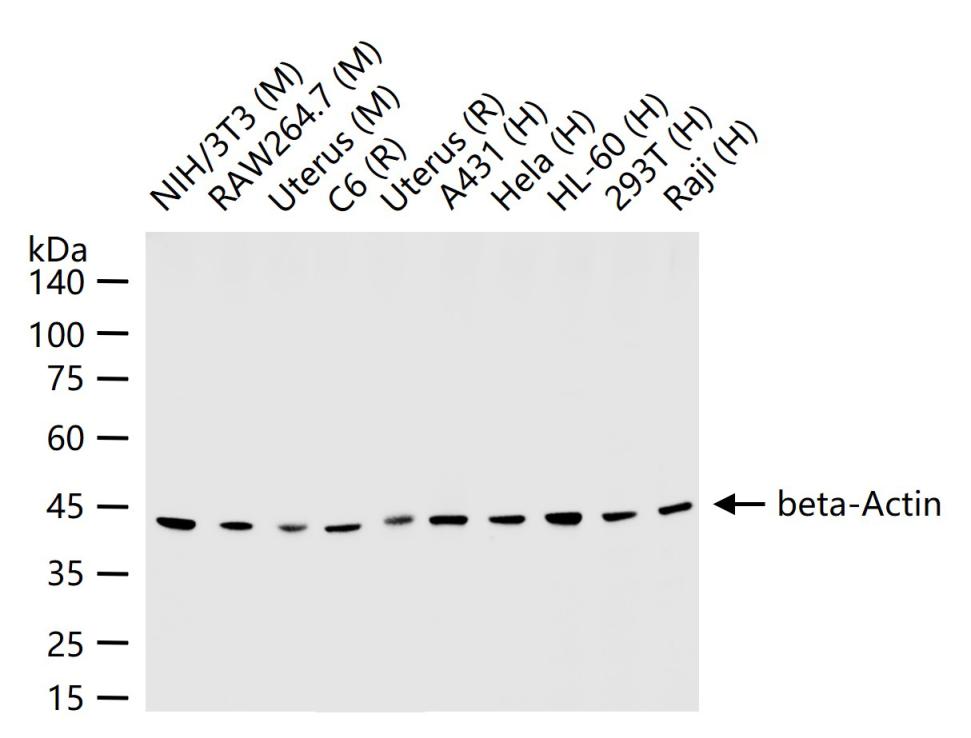
| 产品应用 | WB=1:5000-50000, IHC-P=1:200-1000, IHC-F=1:200-1000, ICC/IF=1:100-500, IF=1:200-1000, Flow-Cyt=1μg/Test Not yet tested in other applications. |
| 交叉反应 | Human, Mouse, Rat (Chicken, Dog, Pig, Rabbit, Sheep, Bee, Fish, GuineaPig, Hamster, Cat) |
| 抗体来源 | Rabbit |
| 免疫原 | Synthetic MAP peptide derived from human beta-Actin |
| 亚型 | IgG |
| 性状 | Liquid |
| 纯化方法 | affinity purified by Protein A |
| 克隆类型 | Polyclonal |
| 理论分子量 | 42 kDa |
| 浓度 | 1mg/ml |
| 储存液 | 0.01M TBS (pH7.4) with 1% BSA, 0.02% Proclin300 and 50% Glycerol. |
| 研究领域 |
Isotype/Loading Controls > Loading Controls > Beta Actin Signal Transduction > Cytoskeleton / ECM > Cytoskeleton > Microfilaments > Actin etc > Actin Tags & Cell Markers > Subcellular Markers > Cytoskeleton > Actin |
| 亚基 | Polymerization of globular actin (G-actin) leads to a structural filament (F-actin) in the form of a two-stranded helix. Each actin can bind to 4 others. Identified in a mRNP granule complex, at least composed of ACTB, ACTN4, DHX9, ERG, HNRNPA1, HNRNPA2B1, HNRNPAB, HNRNPD, HNRNPL, HNRNPR, HNRNPU, HSPA1, HSPA8, IGF2BP1, ILF2, ILF3, NCBP1, NCL, PABPC1, PABPC4, PABPN1, RPLP0, RPS3, RPS3A, RPS4X, RPS8, RPS9, SYNCRIP, TROVE2, YBX1 and untranslated mRNAs. Component of the BAF complex, which includes at least actin (ACTB), ARID1A, ARID1B/BAF250, SMARCA2, SMARCA4/BRG1, ACTL6A/BAF53, ACTL6B/BAF53B, SMARCE1/BAF57 SMARCC1/BAF155, SMARCC2/BAF170, SMARCB1/SNF5/INI1, and one or more of SMARCD1/BAF60A, SMARCD2/BAF60B, or SMARCD3/BAF60C. In muscle cells, the BAF complex also contains DPF3. Found in a complex with XPO6, Ran, ACTB and PFN1. Component of the MLL5-L complex, at least composed of MLL5, STK38, PPP1CA, PPP1CB, PPP1CC, HCFC1, ACTB and OGT. Interacts with XPO6 and EMD. Interacts with ERBB2. |
| 亚细胞定位 | Cytoplasm, cytoskeleton. |
| 组织特异性 | Ubiquitously expressed in all eukaryotic cells. |
| 翻译后修饰 | ISGylated. Oxidation of Met-44 by MICALs (MICAL1, MICAL2 or MICAL3) to form methionine sulfoxide promotes actin filament depolymerization. Methionine sulfoxide is produced stereospecifically, but it is not known whether the (S)-S-oxide or the (R)-S-oxide is produced. |
| 相似性 | Belongs to the actin family. |
| 功能 | Actins are highly conserved proteins that are involved in various types of cell motility and are ubiquitously expressed in all eukaryotic cells. |
| 保存条件 | Shipped at 4℃. Store at -20℃ for one year. Avoid repeated freeze/thaw cycles. |
| 注意事项 | This product as supplied is intended for research use only, not for use in human, therapeutic or diagnostic applications. |
| 背景资料 | Actin is a highly conserved protein and an essential component of cell cytoskeleton and plays an important role in cytoplasmic streaming, cell shape determination, cell division, organelle movement and extension growth. Preferentially expressed in young and expanding tissues, floral organ primordia, developing seeds and emerging inflorescence. Antibodies against plant Actin are useful as loading controls for Western Blotting. |
| 应用 | 推荐稀释比例 |
| {WB} | {1:5000-50000} |
| {IHC-P} | {1:200-1000} |
| {IHC-F} | {1:200-1000} |
| {ICC/IF} | {1:100-500} |
| {IF} | {1:200-1000} |
| {Flow-Cyt} | {1μg/Test} |
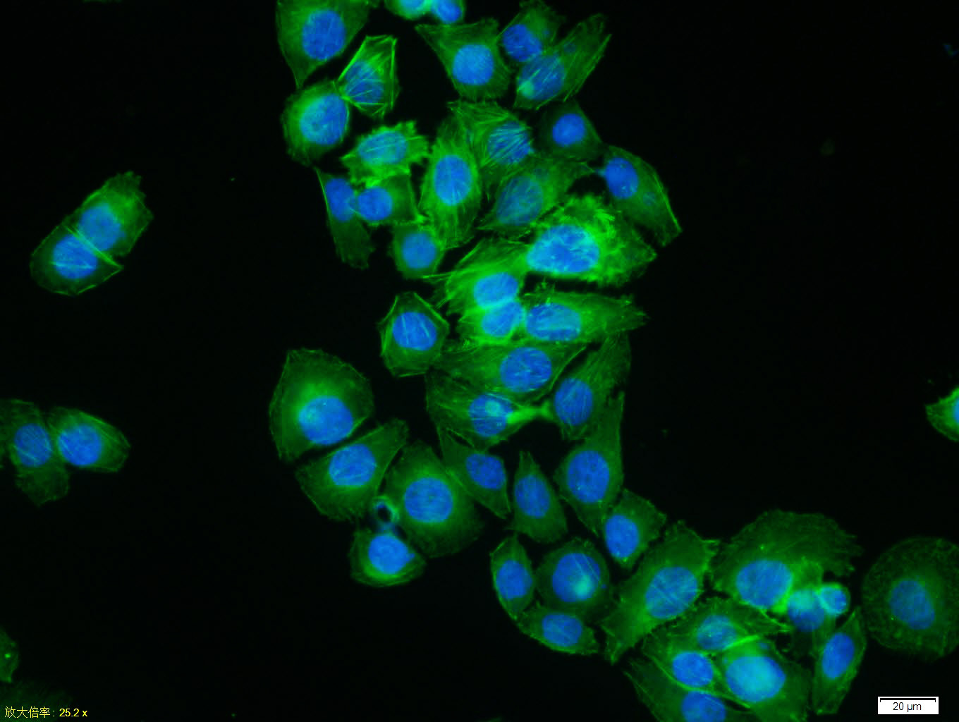
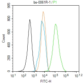
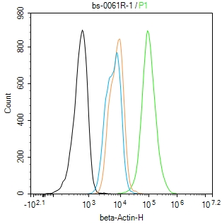
_x000D_ Primary Antibody (green line): Rabbit Anti-beta-Actin (Loading Control) antibody (bs-0061R)
_x000D_ Dilution: 1μg /10^6 cells;
_x000D_ Isotype Control Antibody (orange line): Rabbit IgG .
_x000D_ Secondary Antibody : Goat anti-rabbit IgG-AF488
_x000D_ Dilution: 1μg /test.
_x000D_ Protocol
_x000D_ The cells were fixed with 4% PFA (10min at room temperature)and then permeabilized with 90% ice-cold methanol for 20 min at -20℃. The cells were then incubated in 5%BSA to block non-specific protein-protein interactions for 30 min at room temperature .Cells stained with Primary Antibody for 30 min at room temperature. The secondary antibody used for 40 min at room temperature. Acquisition of 20,000 events was performed._x000D_
风险提示:丁香通仅作为第三方平台,为商家信息发布提供平台空间。用户咨询产品时请注意保护个人信息及财产安全,合理判断,谨慎选购商品,商家和用户对交易行为负责。对于医疗器械类产品,请先查证核实企业经营资质和医疗器械产品注册证情况。
 用户评价
用户评价 暂无用户评价
 文献和实验
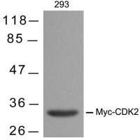
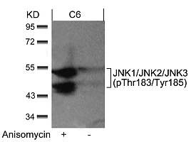
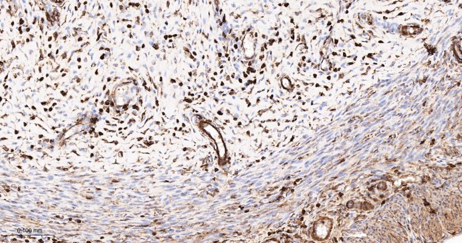
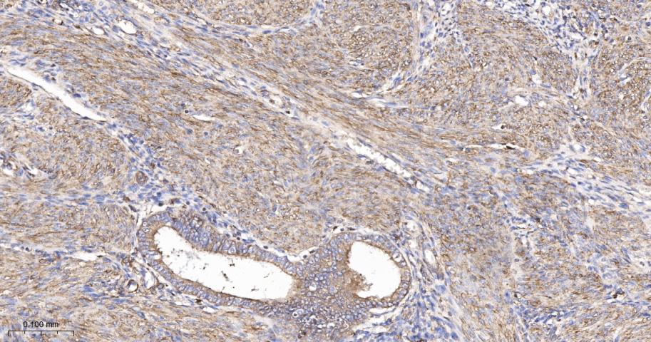
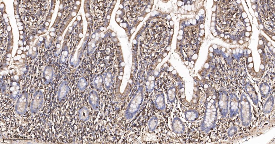
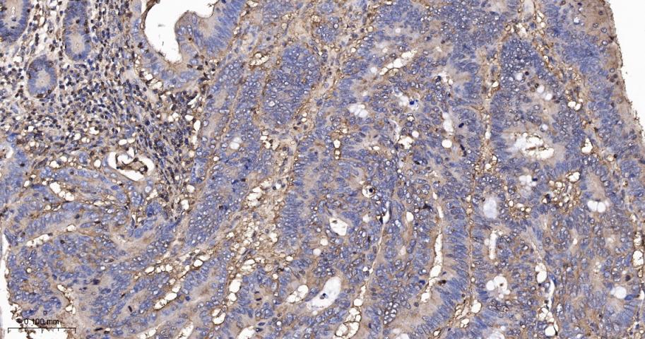
文献和实验[IF={{ preprint }}] {Hui Yang. et al.Identification and Analysis of Key Genes Related to Metabolism in the Brain of Major Depressive Disorder.Research Square.} {Western blot} {Rat}
[IF={{ preprint }}] {Mengyue Yin. et al.Impact of SARS-CoV-2 Infection During Various Pregnancy Trimesters on Maternal and Fetal Outcomes.Research Square.} {Western blot} {Human}
[IF={{ preprint }}] {Ai Fujimoto. et al.Spike protein of the SARS-CoV-2 omicron variant interacts with actin.bioRxiv.} {Western blot} {Mouse}
[IF={{ 9.933 }}] {Xingyi Xu. et al. A Honeycomb-Like Bismuth/Manganese Oxide Nanoparticle with Mutual Reinforcement of Internal and External Response for Triple-Negative Breast Cancer Targeted Therapy. 2021 Jul 23} {WB} {Human}
[IF={{ 9.918 }}] {Jie Hao. et al. Multifunctional miR181a nanoparticles promote highly efficient radiotherapy for rectal cancer. MATER TODAY ADV. 2022 Dec;16:100317} {WB} {Mouse}
duplex PCR amplification, which is the coamplification of a fragment from the region of interest and a control fragment (e.g., the actin gene, or the tubulin gene). This approach allows for estimating relative levels of specific histone modifications
Use of Internaland External Standardsor Reference RNAs for A
levels of the gene under investigation are compared from sample to sample using an internal control to normalize for differences in sample concentration and loading. Ideally, the internal control should be a gene expressed at a constant level
Use of Internal and External Standards or Reference RNAs for
under investigation are compared from sample to sample using an internal control to normalize for differences in sample concentration and loading. Ideally, the internal control should be a gene expressed at a constant level across the sample set. In absolute
 技术资料
技术资料暂无技术资料 索取技术资料

