大家都在搜

手机验证

询价列表

暂时没有已询价产品
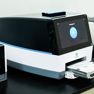
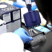
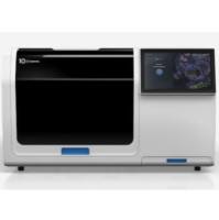
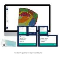
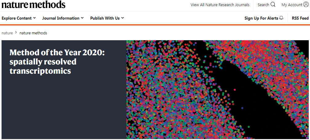
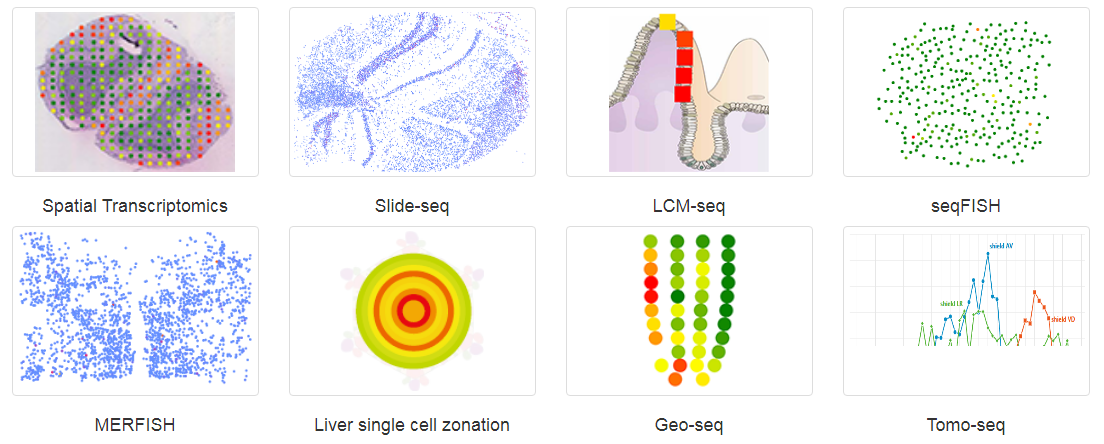
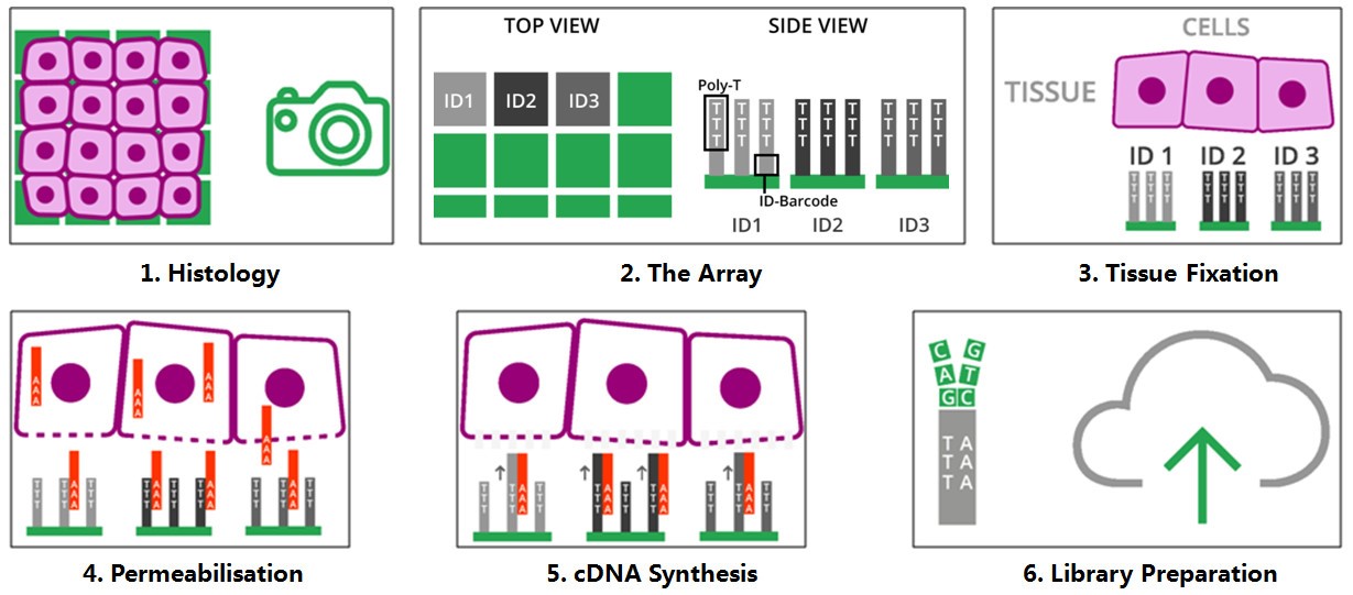
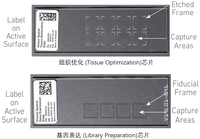
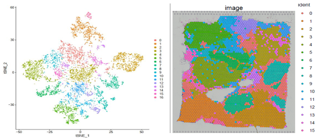
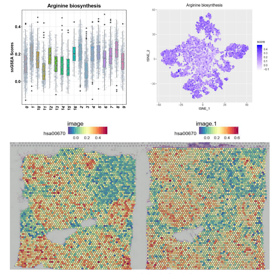
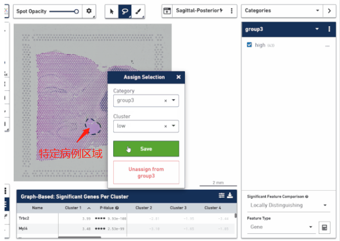
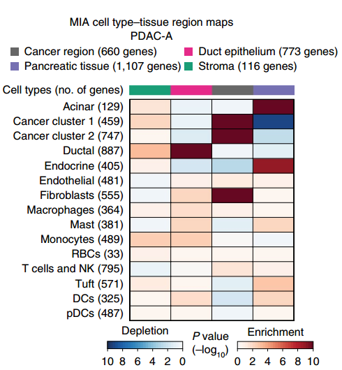
4个空间转录组送2个单细胞(核
)转录组+4个bulk RNA测序
企业认证
伯豪生物
4个空间转录组送2个单细胞(核)转录组+4个bulk RNA测序
以电话询价为准
风险提示:丁香通仅作为第三方平台,为商家信息发布提供平台空间。用户咨询产品时请注意保护个人信息及财产安全,合理判断,谨慎选购商品,商家和用户对交易行为负责。对于医疗器械类产品,请先查证核实企业经营资质和医疗器械产品注册证情况。
 技术资料
技术资料
2023公司简介.pdf 附 (下载 1 次)