


- ¥3500 - 4500
- 欣润生物(NEWGAINBIO)
- 江苏无锡
- IM2021
- 2026年04月25日

企业认证
相关产品推荐更多 >


万千商家帮你免费找货
0 人在求购买到急需产品
- 详细信息
- 文献和实验
- 技术资料
- 英文名:
Immortalized mouse uterine smooth muscle cells
- 库存:
100万
- 供应商:
欣润生物
- 肿瘤类型:
否
- 细胞类型:
永生化
- ATCC Number:
无
- 品系:
ICR
- 组织来源:
子宫
- 相关疾病:
无
- 物种来源:
小鼠
- 免疫类型:
不详
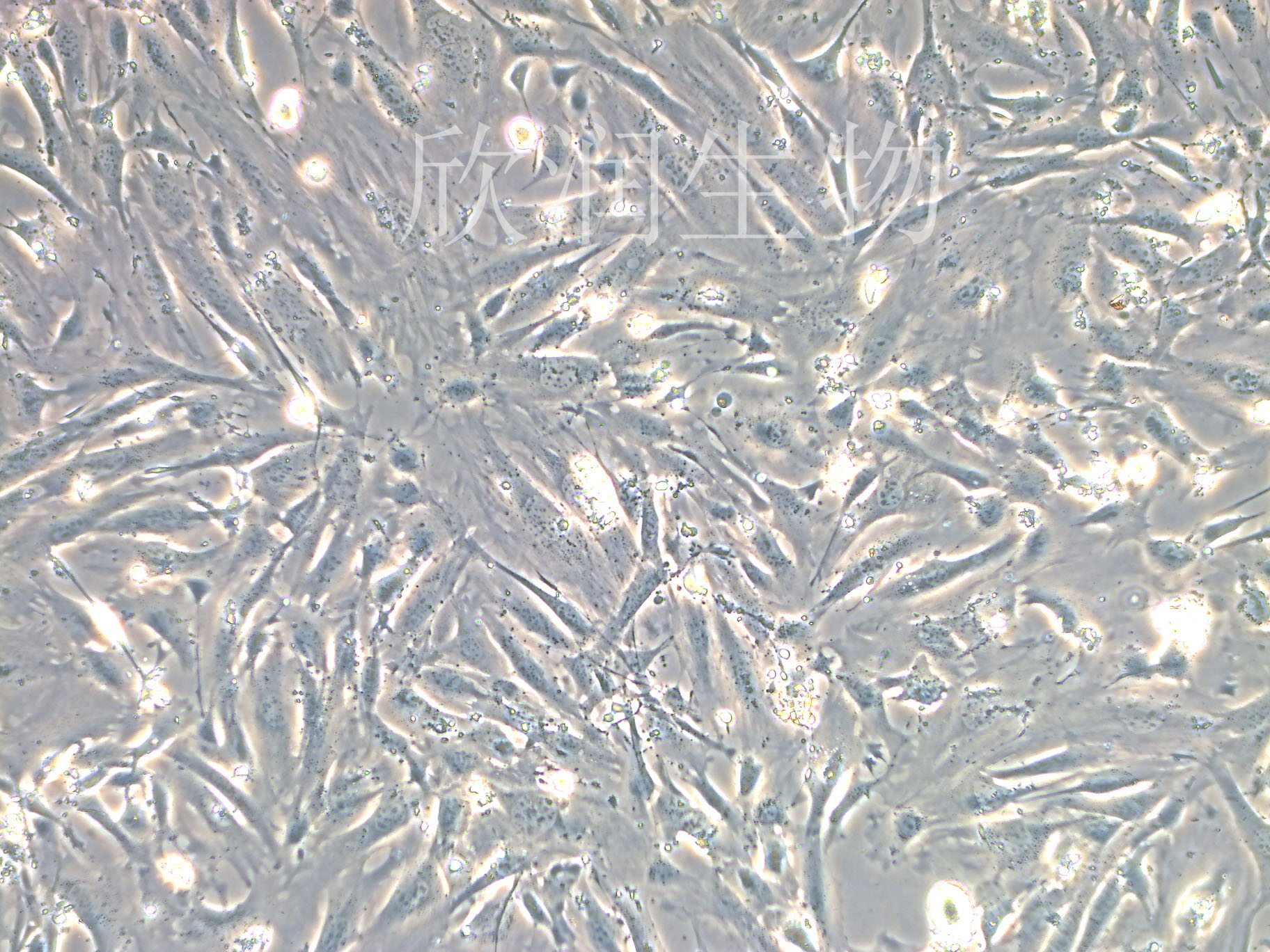
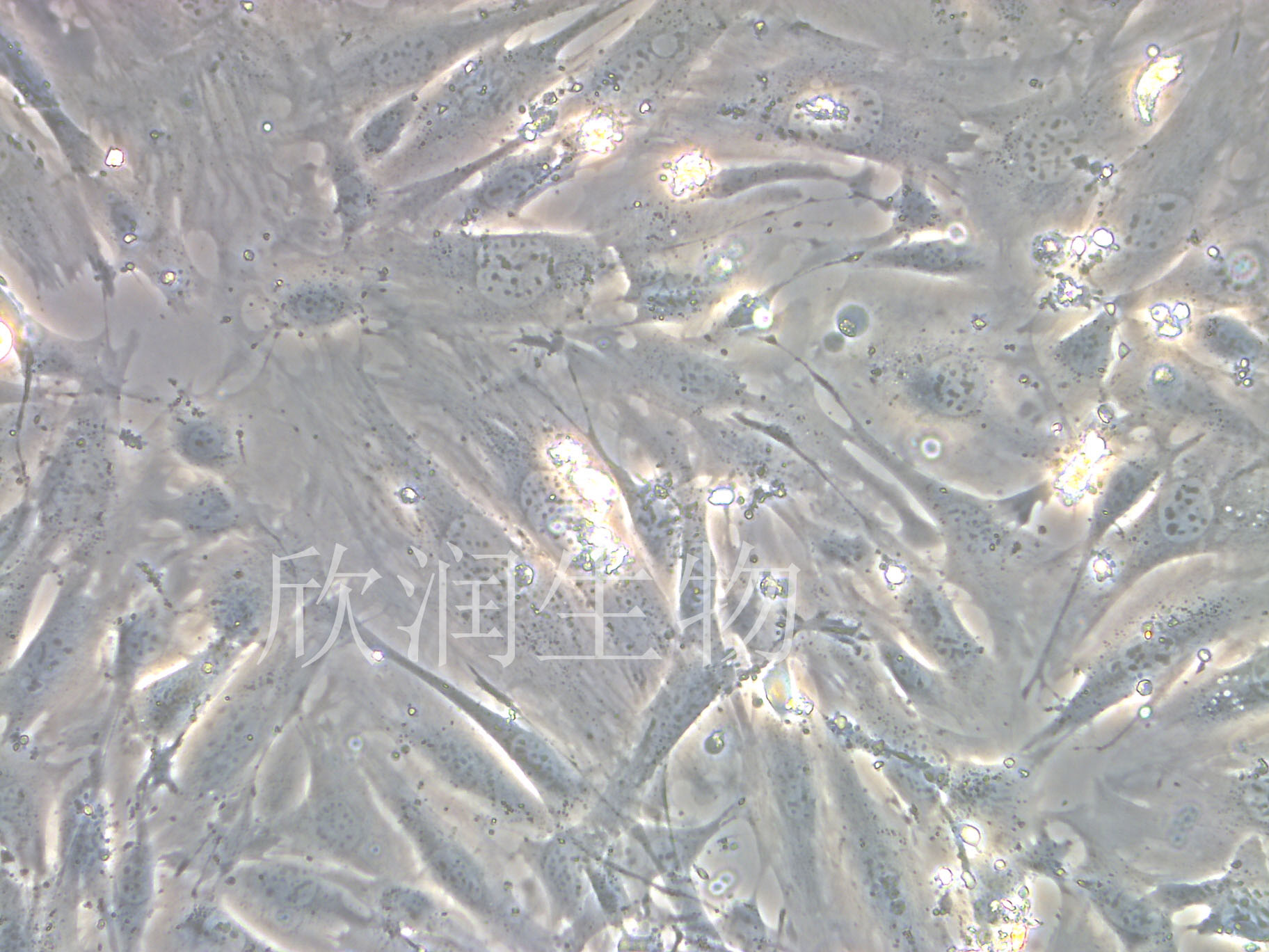
- 细胞形态:
梭形
- 是否是肿瘤细胞:
否
- 器官来源:
子宫
- 运输方式:
常温
- 年限:
/
- 生长状态:
贴壁生长
- 规格:
T25方瓶
永生化小鼠子宫平滑肌细胞简介:
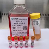
产品名称:小鼠子宫平滑肌细胞系
产品描述:小鼠子宫是小鼠的雌性生殖器官的一部分,是小鼠胎儿或幼体发育生长的场所。子宫肌层比较厚,由成束或成片的平滑肌组成,肌束间以结缔组织分隔。子宫平滑肌具有收缩功能,收缩受激素的调节,其收缩活动有助于精子向输卵管运送、经血排出以及胎儿娩出。人子宫平滑肌细胞系、永生化人子宫平滑肌细胞的分裂增殖还受性腺激素的影响。产品货号:IM2011
产品类型: 有限细胞系
传代能力: 传代30代左右
产品形态: 成纤维细胞样
培养基:永生化小鼠子宫平滑肌细胞专用完全培养基
支原体检测:呈阴性
产品培养条件:37℃,5%CO2
发货方式:T25方瓶发货
货期:1周左右时间
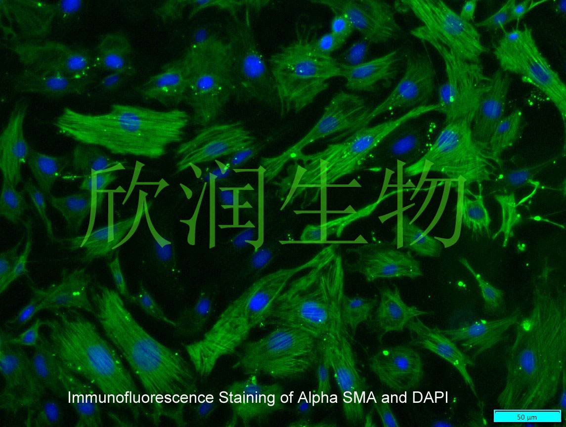
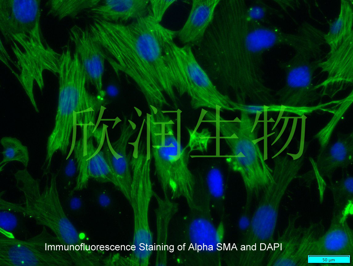
细胞鉴定:a-SMA免疫荧光呈阳性
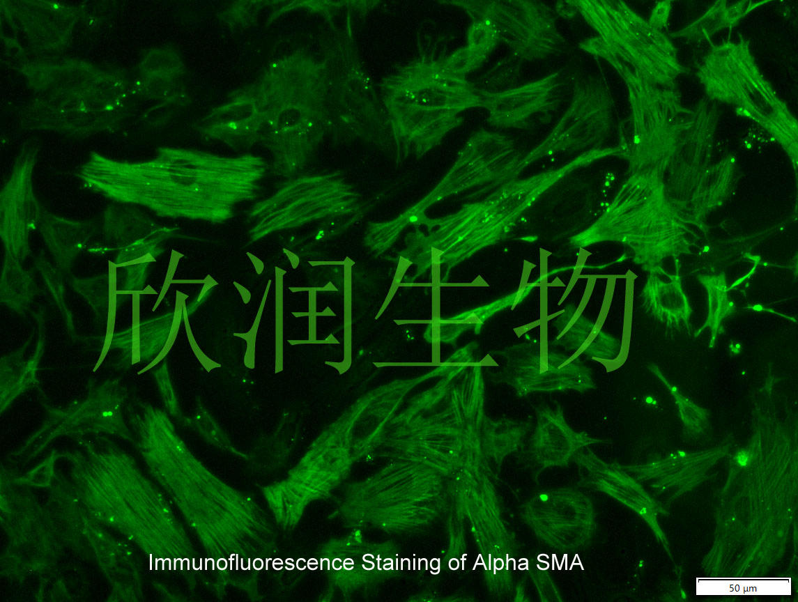
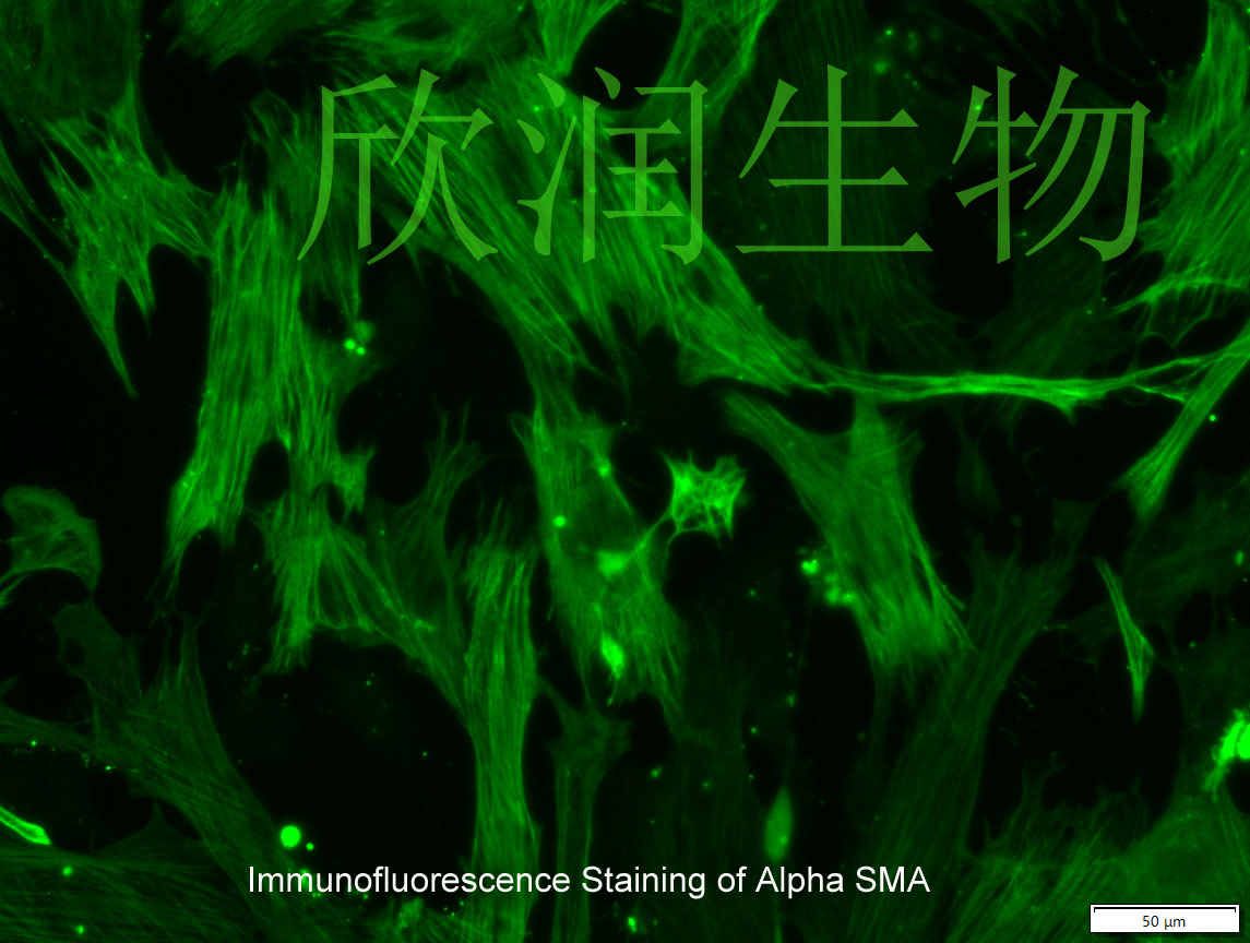
Alpha SMA免疫荧光染色鉴定


Expression of Mel-CAM in implantation site intermediate trophoblastic cell line, IST-1, limits its migration on uterine smooth muscle cells.
An immortalized implantation site intermediate trophoblastic cell line, IST-1, was established from a human placenta of 7 weeks gestation. IST-1 cells phenotypically resembled the implantation site intermediate trophoblastic cells in situ and expressed Mel-CAM (MUC 18 or CD146). Mel-CAM is a cell adhesion molecule belonging to the immunoglobulin gene superfamily. It is involved in heterophilic cell-cell adhesion and plays a role in several biological processes including tumor progression. We have previously shown that Mel-CAM was highly expressed in the intermediate (extravillous) trophoblast in the human implantation site. In this study we determined the function of Mel-CAM in the interaction of trophoblast and uterine smooth muscle in the implantation site. IST-1 cells failed to adhere to immobilized recombinant Mel-CAM in solid phase whereas the uterine smooth muscle cells did. The presence of the putative Mel-CAM ligand in smooth muscle cells was further supported by the finding that Mel-CAM-transfected but not the mock-transfected U937 leukemia
风险提示:丁香通仅作为第三方平台,为商家信息发布提供平台空间。用户咨询产品时请注意保护个人信息及财产安全,合理判断,谨慎选购商品,商家和用户对交易行为负责。对于医疗器械类产品,请先查证核实企业经营资质和医疗器械产品注册证情况。
 文献和实验
文献和实验Chicken intestinal epithelial cells were obtained from NEWGAINBIO company. Cells were cultured on 37℃, with 5% CO2, in the Ham’s F-12 Nutrient (DMEM/12) that contained the following supplementations: fetal bovine serum (5%), in-sulin (5 µg/mL), transferrin (5 µg/mL), selenium (5 ng/mL), epidermal growth factor (5 ng/mL) and penicillin-streptomycin (100–100 U/mL) for cell culturing (full DMEM/12). Experiments were performed with chicken intestinal epithelial cells and working solutions were prepared with plain DMEM/12 without supplementation. For the investigations, cells were seeded onto 96-well, 24-well or 6-well polystyrene cell culture plates.
Primary hVICs (passage 2) were cultured to 50–60% confluence and infected with pGMLV-SV40T-puro lentivirus (NewgainBio, Wuxi, China) at a multiplicity of infection of 80 supplemented with 5 µg/mL polybrene (Sigma-Aldrich, Buchs, Switzerland).
Tissue was cultured until cells became visible around the tissue, and when the fusion reached 90% (FIGURE 1A) §ask ¦lled with the prepared culturing medium was sent to the company for further immortalisation. Cell immortalisation was done for cell stability and longer-term use. Immortalised cells were cultured with 10% FBS and 1% PS in the DMEM medium. After the cells multiplied and merged, they were routinely passed and grown ( NEWGAINBIO Inc. Wuxi, Jiangsu, China) (FIGURE 1B-C).
Mouse primary cultured renal vascular ECs and VSMCs were obtained from Newgainbio company, which were tested by Factor VIII and α-smooth muscle actin (α-SMA), the marker of ECs and VSMCs. RNeasy Mini Kit was used for RNA extraction, and the above protocols were repeated.
Porcine primary colon epithelial cells (Newgainbio company, Wuxi,China) were cultured in Dulbecco's Modified Eagle's Medium (Solarbio, Beijing, China) containing 10 % fetal bovine serum (BioInd, Kiryat shmona, Lsrael) at 37 ◦C and 5 % CO2 humidity.
3K的下游靶点。 刚刚接触这方面的信号通路,望多指教,谢谢! yhwangcams 呵呵,好多情况下得出的结论都可能源于不同的细胞系,不同的处理方法,等等。 各个信号通路间会有一些“交流”(crosstalk)。我的建议是先从Nature, Cell等上找一些大人物写的综述看看。基本了解后,再看相关细胞里(如平滑肌细胞等)的研究。自己能做出什么结果,只要可信,就可以得出相关结论。 本文由丁香园论坛提供,想了解更多有用的、有意
主要有两类:神经嵴干细胞(neuralcreststemcell,NC-SC)和中枢神经干细胞(CNS-SC)。NCSC为外周神经干细胞(PNS-SC),既可发育为外周神经细胞、神经内分泌细胞和Schwann氏细胞,也能分化为色素细胞(pigmented cell)和平滑肌细胞等。NSC一般是指存在于脑部的中枢神经干细胞(CNS-SC),其子代细胞能分化成为神经系统的大部分细胞。以往认为,中枢神经系统的神经元在出生前或出生后不久,就失去再生能力。但近年的一些研究表明,成年哺乳
、物理、病毒等方法)诱导细胞发生转化,使其倍增时间减少,永生化或生命期延长,也是一个努力方向。 但是要建立适于组织工程需要的种子细胞,需要解决以下问题:①增加细胞的增殖能力;②延长细胞的生命期;③提高细胞的分泌能力;④优选不同组织来源的同一功能的最佳细胞;⑤建立标准细胞系,使研究工作有更好的可比性和科学性;⑥同种异体与异种移植的免疫学;⑦细胞与人工细胞外基质的相互作用及影响因素。 采用同种异体细胞来源,在目前仅对少数组织细胞(如软骨组织的组织工程培养)有望获得
 技术资料
技术资料
