The Effects of Calcium-Free Seawater on the Development of Sea Urchin Embryos
互联网
Objective
The purpose of this experiment is to determine the effects of calcium-free seawater on the development of sea urchin embryos. Embryos were placed in calcium-free sea after fertilization and at the initiation of gastrulation
Introduction
Calcium is necessary in two processes shortly following fertilization. One process, the acrosomal reaction, is the fusion of the acrosomal vesicle and the sperm plasma membrane, which results in the extension of the acrosomal process. The acrosomal reaction is initiated by a fucose-containing polysaccharide within the egg jelly, which binds to the sperm and allows calcium to enter the sperm head. The second mechanism is the cortical granule reaction. Upon fertilization, the calcium ion concentration of the egg increases greatly, which causes the cortical granule membranes to fuse with the egg plasma membrane, releasing their contents. A wave of cortical, released from the endoplasmic reticulum, begins at the point of fertilization and travels completely around the egg. The calcium ions needed for these two mechanisms are not a result of an influx of calcium, but come from within the egg itself. Therefore withdrawing calcium during fertilization would not alter these processes.
Later in development, calcium may play a role in cell adhesion. The major cell adhesion molecules are known as cadherins. Cadherins are calcium-dependent adhesion molecules. Cadherins are critical in establishing and maintaining intercellular connections, and appear to be crucial to the spatial segregation of cell types and to the organization of animal form. Calcium ions are needed for the adhesion of the same cadherin molecules between cells.
The sea urchin embryo provides a powerful model system for the study of morphogenesis and cadherin activity. In a study performed by Jeffrey Miller and David McClay, three developmental events where show to be directly related to cadherin molecules: 1) the acquisition of cell polarity in cleavage-stage blastomeres; 2) the epithelial- mesenchymal conversion of epithelial cells to mesodermal derivatives; and 3) the convergent extension movements involved in constructing the archenteron. Their data provided new insight into the function of cadherins during various morphogenetic events and provided building blocks for future experiments exploring cadherin localization and adhesive activity during development.

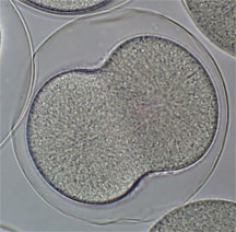
Figure: Early sea urchin embryo performing
first cleavage
Procedure
with calcium-free seawater.
document transfer to calcium-free seawater.
order to remove as much calcium as possible.
embryos.
as experimental embryo for comparison.
Results
The embryos were observed after a 24 hours time period. There was strong evidence to show the calcium is a major contributor in the development of the sea urchin embryos. The control embryos developed normally and at time of observation were performing gastrulation (See Figure 1 ). The embryos, which were placed in calcium-free sea water immediately following fertilization, began development normally and were similar to the control embryos at the 7-hour stage (See Figure 2 and 3 ). But when they were observed at the 20-hour stage the embryos had not continued through gastrulation ( See Figure 4 ). The embryos appeared to stop development soon after the 7-hour stage. The embryos, which were placed in calcium-free sea water after 7 hours of development, showed very different results ( See Figure 5 ). The embryos had died and no longer maintained their shape. It appeared as if the cells were no longer adhered together. Based on these results, major events during development are dependent on the presence of calcium. In the future, further developments can be made involving the importance of cadherin molecules in embryogenesis.