• 我要登录|
  • 免费注册
    |
  • 我的丁香通
    • 企业机构:
    • 成为企业机构
    • 个人用户:
    • 个人中心
  • 移动端
    移动端
丁香通 logo丁香实验_LOGO
搜实验

    大家都在搜

      大家都在搜

        0 人通过求购买到了急需的产品
        免费发布求购
        发布求购
        点赞
        收藏
        wx-share
        分享

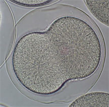
        Effect of UV Treatment on Early Development of Sea Urchin Embryos

        互联网

        912


        Objective:
        To test the effects of UV radiation at 254nm on Sea Urchin embryo development after radiation at the 2-cell stage.

        Background:
        Sea Urchins exhibit radial holoblastic cleavage. The first and second cleavages are perpendicular to one another

        and go through the animal and vegetal poles. The subsequent cleavages separate the cells into mesomeres,

        macromeres, and micromeres. Sea Urchins undergo formation of the blastula and gastrulation before developing into a

        mature embryo.

        UV light is harmful to many organisms just as it is harmful to humans. Many eggs of marine animals including sea

        urchins have a high concentration of mycosporine amino acids that protect them from UV damage. Mycosporine amino

        acids are obtained through the diet of the adult and are stored in the egg. However, there is fear that the amphibians are

        declining because of the increased UV-B reaching the earth's surface. UV-A and UV-B reach the earth's surface. UV-B is

        increasingly hitting the earth's surface because of the depleting ozone layer. One of the more common effects of UV-B is

        adjacent thymidines condensing into cyclobutane pyrimidine dimers (CBPDs) which impede DNA replication and

        transcription. UV-C (200-280nm) is the most harmful type of UV to organisms, however, due to oxygen in the atmosphere


        absorbing the UV rays, UV-C never reaches the Earth's surface. The effects of ozone depletion are likely to be most

        devastating to organisms that are exposed to sunlight during their developmental stages. The sea urchin embryos are

        transparent and poorly protected against UV. Sea urchins are exposed to UV radiation during all stages of their lifecycle.

        Sea Urchins like many organisms have DNA repair enzymes, photolyases. UV radiation causes direct damage to proteins

        and DNA by creating the formation of pyrimidine dimers and crosslinks between DNA and protein. Consequences of

        these mutations are delayed cell divisions, DNA damage, developmental delays, and abnormalities. UV treatment can

        cause immediate effects by damaging microtubules and long term effect by damaging RNA and DNA.

        Experimental Design:

        gametes in artificial seawater ( ASW ) 3 times

        2) Add 1ml of diluted sperm to the gametes in ASW ( Fertilize gamates )

        3) Wait until more than 75% of the embryos undergo first cleavage (check for cleavage under compound microscope periodically) See Figure 1.

        4) Split the embryos into 4 open dishes labeled; 1min UV , 2 min UV, 5 min UV, Control

        5) Place the open dish with embryos labeled 1 min UV 3 1/2 inches under the hand held UV lamp and turn on the short wave lamp for 1 minute while wearing protective goggles

        6) Repeat for the two dishes labeled, 2 min UV, 5 min UV for times indicated

        7) Wait for the second cleavage to occur, take pictures with digital camera while under the microscope of the control embryos and the three experimental groups of embryos.

        8) Wait for the control embryos to hatch

        9) Come in the next morning look under microscope and take pictures of all 4 groups of embryos to compare stages of development and percentage of normally developed cells

        Figure 1. Sea Urchin embryos during first cleavage when radiated with UV-C.

        Results and Discussion:

        At second cleavage, all the eggs that were radiated with UV for one minute looked normal. At second cleavage,

        most of the eggs that were radiated with UV for two minutes looked normal. A couple of eggs looked like Figure 2. They

        had a cytoplasmic blip. At second cleavage, majority of the eggs that were radiated with UV for five minutes looked

        normal. A handful of eggs looked the same as Figure 2. The UV did not seem to damage the embryos immediately; most

        of the second cleavages were normal for all eggs in all the experimental exposure times.

        In the second part of the experiment, the embryos were observed after gastrulation. At that point the archenteron

        is visible, secondary mesenchyme cells are visible, and cells organized themselves into spicules. All the control eggs

        were hatched from the fertilization envelope and were ciliated as in Figure 3. All the embryos that were radiated with UV

        for one minute had blastula and cilia and primary mesenchyme cells but no vegetal plate or vagination was observed.

        This is seen in Figure 4. All the embryos that were radiated with UV for two minutes looked the same as Figure 4 except

        one normal embryo was found. All the cells that were radiated with UV for five minutes looked the same as Figure 4. All

        the abnormal embryos had abnormally large amounts of cells in the blastocoel. All the cells radiated with UV-C seem

        delayed in development or dead at the beginning of development of the vegetal plate. Most embryos did not undergo

        gastrulation. The UV-C damages the embryos somewhere between the second cleavage and gastrulation. The amount of

        exposure time to UV-C does not seem to determine the amount of development deformities. All the experimental

        exposure times to UV-C embryos stopped developing after the formation of the blastula but before gastrulation. This

        suggests that the mycosporine amino acids do not help as much with protection against UV-C as they do with UV-B.

        Figure 2. Sea Urchin embryos radiated with UV-C at second cleavage with a cytoplasmic blip

        Figure 3. Control Sea Urchin embryos hatched from the fertilization envelope,ciliated, and undergoing gastrulation.

        Figure 4. UV radiated Sea Urchin embryos delayed development and excess cells in the blastocoel


        ad image
        提问
        扫一扫
        丁香实验小程序二维码
        实验小助手
        丁香实验公众号二维码
        扫码领资料
        反馈
        TOP
        打开小程序