Preparation of Recombinant Protein Spotted Arrays for Proteome‐Wide Identification of Kinase Targets
互联网
- Abstract
- Table of Contents
- Materials
- Figures
- Literature Cited
Abstract
Protein microarrays allow unique approaches for interrogating global protein interaction networks. Protein arrays can be divided into two categories: antibody arrays and functional protein arrays. Antibody arrays consist of various antibodies and are appropriate for profiling protein abundance and modifications. Functional full?length protein arrays employ full?length proteins with various post?translational modifications. A key advantage of the latter is rapid parallel processing of large number of proteins for studying highly controlled biochemical activities, protein?protein interactions, protein?nucleic acid interactions, and protein?small molecule interactions. This unit presents a protocol for constructing functional yeast protein microarrays for global kinase substrate identification. This approach enables the rapid determination of protein interaction networks in yeast on a proteome?wide level. The same methodology can be readily applied to higher eukaryotic systems with careful consideration of overexpression strategy. Curr. Protoc. Protein Sci. 72:27.4.1?27.4.14. © 2013 by John Wiley & Sons, Inc.
Keywords: protein array; post?translation; phosphorylation; kinase
Table of Contents
- Introduction
- Strategic Planning
- Basic Protocol 1: Protein Induction and Purification of Proteins for Printing
- Basic Protocol 2: Printing of Proteome Microarrays
- Basic Protocol 3: Perform Radioactive In Vitro Kinase Assay on a Protein Microarray
- Reagents and Solutions
- Commentary
- Literature Cited
- Figures
- Tables
Materials
Basic Protocol 1: Protein Induction and Purification of Proteins for Printing
Materials
Basic Protocol 2: Printing of Proteome Microarrays
Materials
Basic Protocol 3: Perform Radioactive In Vitro Kinase Assay on a Protein Microarray
Materials
|
Figures
-
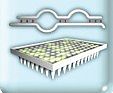
Figure 27.4.1 Schematic diagram of overall protocol. Yeast clones containing cDNA constructs are plated on selection medium. Each clone is grown and protein expression is induced. Pellets of each clone are transferred into 96‐well boxes. Fusion proteins are purified and transferred to 96‐well titer plates for printing on slides. In the upper right, the kinase‐treated block shows spots, which indicate phosphorylation of printed proteins with radioactive phosphate. Spots in the corner rectangles are positive controls. View Image
Videos
Literature Cited
| Balboni, I., Limb, C., Tenenbaum, J.D., and Utz, P.J. 2008. Evaluation of microarray surfaces and arraying parameters for autoantibody profiling. Proteomics 8:3443‐3449. | |
| Brizuela, L., Braun, P., and LaBaer, J. 2001. FLEXGene repository: From sequenced genomes to gene repositories for high‐throughput functional biology and proteomics. Mol. Biochem. Parasitol. 118:155‐165. | |
| Bussow, K., Nordhoff, E., Lubbert, C., Lehrach, H., and Walter, G. 2000. A human cDNA library for high‐throughput protein expression screening. Genomics 65:1‐8. | |
| Cheung, K.H., Hager, J., Nelson, K., White, K., Li, Y., Snyder, M., Williams, K., and Miller, P. 2002. A dynamic approach to mapping coordinates between microplates and microarrays. J. Biomed. Informatics 35:306‐312. | |
| Fasolo, J., Sboner, A., Sun, M.G., Yu, H., Chen, R., Sharon, D., Kim, P.M., Gerstein, M., and Snyder, M. 2011. Diverse protein kinase interactions identified by protein microarrays reveal novel connections between cellular processes. Genes Dev. 25:767‐778. | |
| Gelperin, D.M., White, M.A., Wilkinson, M.L., Kon, Y., Kung, L.A., Wise, K.J., Lopez‐Hoyo, N., Jiang, L., Piccirillo, S., Yu, H., Gerstein, M., Dumont, M.E., Phizicky, E.M., Snyder, M., and Grayhack, E.J. 2005. Biochemical and genetic analysis of the yeast proteome with a movable ORF collection. Genes Dev. 19:2816‐2826. | |
| Graslund, S., Nordlund, P., Weigelt, J., Hallberg, B.M., Bray, J., Gileadi, O., Knapp, S., Oppermann, U., Arrowsmith, C., Hui, R., Ming, J., dhe‐Paganon, S., Park, H.W., Savchenko, A., Yee, A., Edwards, A., Vincentelli, R., Cambillau, C., Kim, R., Kim, S.H., Rao, Z., Shi, Y., Terwilliger, T.C., Kim, C.Y., Hung, L.W., Waldo, G.S., Peleg, Y., Albeck, S., Unger, T., Dym, O., Prilusky, J., Sussman, J.L., Stevens, R.C., Lesley, S.A., Wilson, I.A., Joachimiak, A., Collart, F., Dementieva, I., Donnelly, M.I., Eschenfeldt, W.H., Kim, Y., Stols, L., Wu, R., Zhou, M., Burley, S.K., Emtage, J.S., Sauder, J.M., Thompson, D., Bain, K., Luz, J., Gheyi, T., Zhang, F., Atwell, S., Almo, S.C., Bonanno, J.B., Fiser, A., Swaminathan, S., Studier, F.W., Chance, M.R., Sali, A., Acton, T.B., Xiao, R., Zhao, L., Ma, L.C., Hunt, J.F., Tong, L., Cunningham, K., Inouye, M., Anderson, S., Janjua, H., Shastry, R., Ho, C.K., Wang, D., Wang, H., Jiang, M., Montelione, G.T., Stuart, D.I., Owens, R.J., Daenke, S., Schutz, A., Heinemann, U., Yokoyama, S., Bussow, K., and Gunsalus, K.C. 2008. Protein production and purification. Nat. Methods 5:135‐146. | |
| Hu, S., Xie, Z., Onishi, A., Yu, X., Jiang, L., Lin, J., Rho, H.S., Woodard, C., Wang, H., Jeong, J.S., Long, S., He, X., Wade, H., Blackshaw, S., Qian, J., and Zhu, H. 2009. Profiling the human protein‐DNA interactome reveals ERK2 as a transcriptional repressor of interferon signaling. Cell 139:610‐622. | |
| Hudson, J.R., Jr., Dawson, E.P., Rushing, K.L., Jackson, C.H., Lockshon, D., Conover, D., Lanciault, C., Harris, J.R., Simmons, S.J., Rothstein, R., and Fields, S. 1997. The complete set of predicted genes from Saccharomyces cerevisiae in a readily usable form. Genome Res. 7:1169‐1173. | |
| Lennon, G., Auffray, C., Polymeropoulos, M., and Soares, M.B. 1996. The I.M.A.G.E. Consortium: an integrated molecular analysis of genomes and their expression. Genomics 33:151‐152. | |
| MacBeath, G. and Schreiber, S.L. 2000. Printing proteins as microarrays for high‐throughput function determination. Science 289:1760‐1763. | |
| Meng, L., Michaud, G.A., Merkel, J.S., Zhou, F., Huang, J., Mattoon, D.R., and Schweitzer, B. 2008. Protein kinase substrate identification on functional protein arrays. BMC Biotechnol. 8:22. | |
| Popescu, S.C., Popescu, G.V., Bachan, S., Zhang, Z., Seay, M., Gerstein, M., Snyder, M., and Dinesh‐Kumar, S.P. 2007. Differential binding of calmodulin‐related proteins to their targets revealed through high‐density Arabidopsis protein microarrays. Proc. Natl. Acad. Sci. U.S.A. 104:4730‐4735. | |
| Ptacek, J., Devgan, G., Michaud, G., Zhu, H., Zhu, X., Fasolo, J., Guo, H., Jona, G., Breitkreutz, A., Sopko, R., McCartney, R.R., Schmidt, M.C., Rachidi, N., Lee, S.J., Mah, A.S., Meng, L., Stark, M.J., Stern, D.F., De Virgilio, C., Tyers, M., Andrews, B., Gerstein, M., Schweitzer, B., Predki, P.F., and Snyder, M. 2005. Global analysis of protein phosphorylation in yeast. Nature 438:679‐684. | |
| Reboul, J., Vaglio, P., Rual, J.F., Lamesch, P., Martinez, M., Armstrong, C.M., Li, S., Jacotot, L., Bertin, N., Janky, R., Moore, T., Hudson, J.R., Jr., Hartley, J.L., Brasch, M.A., Vandenhaute, J., Boulton, S., Endress, G.A., Jenna, S., Chevet, E., Papasotiropoulos, V., Tolias, P.P., Ptacek, J., Snyder, M., Huang, R., Chance, M.R., Lee, H., Doucette‐Stamm, L., Hill, D.E., and Vidal, M. 2003. C. elegans ORFeome version 1.1: Experimental verification of the genome annotation and resource for proteome‐scale protein expression. Nat. Genet. 34:35‐41. | |
| Schweitzer, B. and Kingsmore, S.F. 2002. Measuring proteins on microarrays. Curr. Opin. Biotechnol. 13:14‐19. | |
| Strausberg, R.L., Feingold, E.A., Klausner, R.D., and Collins, F.S. 1999. The mammalian gene collection. Science 286:455‐457. | |
| Strausberg, R.L., Feingold, E.A., Grouse, L.H., Derge, J.G., Klausner, R.D., Collins, F.S., Wagner, L., Shenmen, C.M., Schuler, G.D., Altschul, S.F., Zeeberg, B., Buetow, K.H., Schaefer, C.F., Bhat, N.K., Hopkins, R.F., Jordan, H., Moore, T., Max, S.I., Wang, J., Hsieh, F., Diatchenko, L., Marusina, K., Farmer, A.A., Rubin, G.M., Hong, L., Stapleton, M., Soares, M.B., Bonaldo, M.F., Casavant, T.L., Scheetz, T.E., Brownstein, M.J., Usdin, T.B., Toshiyuki, S., Carninci, P., Prange, C., Raha, S.S., Loquellano, N.A., Peters, G.J., Abramson, R.D., Mullahy, S.J., Bosak, S.A., McEwan, P.J., McKernan, K.J., Malek, J.A., Gunaratne, P.H., Richards, S., Worley, K.C., Hale, S., Garcia, A.M., Gay, L.J., Hulyk, S.W., Villalon, D.K., Muzny, D.M., Sodergren, E.J., Lu, X., Gibbs, R.A., Fahey, J., Helton, E., Ketteman, M., Madan, A., Rodrigues, S., Sanchez, A., Whiting, M., Madan, A., Young, A.C., Shevchenko, Y., Bouffard, G.G., Blakesley, R.W., Touchman, J.W., Green, E.D., Dickson, M.C., Rodriguez, A.C., Grimwood, J., Schmutz, J., Myers, R.M., Butterfield, Y.S., Krzywinski, M.I., Skalska, U., Smailus, D.E., Schnerch, A., Schein, J.E., Jones, S.J., and Marra, M.A. 2002. Generation and initial analysis of more than 15,000 full‐length human and mouse cDNA sequences. Proc. Natl. Acad. Sci. U.S.A. 99:16899‐16903. | |
| Tabakman, S.M., Lau, L., Robinson, J.T., Price, J., Sherlock, S.P., Wang, H., Zhang, B., Chen, Z., Tangsombatvisit, S., Jarrell, J.A., Utz, P.J., and Dai, H. 2011. Plasmonic substrates for multiplexed protein microarrays with femtomolar sensitivity and broad dynamic range. Nat. Communications 2:466. | |
| Tsvetanova, N.G., Klass, D.M., Salzman, J., and Brown, P.O. 2010. Proteome‐wide search reveals unexpected RNA‐binding proteins in Saccharomyces cerevisiae. PLoS One 5:e12671. | |
| Zhu, H., Bilgin, M., Bangham, R., Hall, D., Casamayor, A., Bertone, P., Lan, N., Jansen, R., Bidlingmaier, S., Houfek, T., Mitchell, T., Miller, P., Dean, R.A., Gerstein, M., and Snyder, M. 2001. Global analysis of protein activities using proteome chips. Science 293:2101‐2105. | |
| Zhu, X., Gerstein, M., and Snyder, M. 2006. ProCAT: a data analysis approach for protein microarrays. Genome Biol. 7:R110. | |
| Internet Resources | |
| http://www.invitrogen.com/site/us/en/home/LINNEA‐Online‐Guides/LINNEA‐Guide‐to‐Clones/Ultimate‐ORF‐Clones.html | |
| Invitrogen's Ultimate ORF clone information Web site has more details on available cDNA collections and expression strategy using GATEWAY technology | |
| http://www.invitrogen.com/site/us/en/home/Products‐and‐Services/Applications/Protein‐Expression‐and‐Analysis/Biomarker‐Discovery/ProtoArray/Resources/Data‐Analysis.html | |
| Information about Invitrogen's Prospector software to analyze protein array data. | |
| http://www.openbiosystems.com | |
| Web site for OpenBiosystems, a distributor of many of cDNA collection now owned by Thermo. | |
| http://www.origene.com | |
| Web site for Origene. It houses expression validated cDNA clones. | |
| http://dnasu.asu.edu/DNASU | |
| Web site for the ASU biodesign institute. An academic resource for obtaining various mammalian cDNA collections and expression vector plasmid for nominal fee. | |
| http://ymd.med.yale.edu/kei‐cgi/kc_mac_dev8.pl | |
| MicroArray Convolutor. Web tool for generating gal file for microarray analysis. |