


- 询价
- 2026年05月29日

企业认证


万千商家帮你免费找货
0 人在求购买到急需产品
- 详细信息
- 询价记录
- 文献和实验
- 技术资料
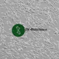
- 器官来源:
肺
- 物种来源:
人
- 是否是肿瘤细胞:
1
- 年限:
58 years
- 相关疾病:
肿瘤
- ATCC Number:
CCL-185™
- 细胞形态:
上皮样
- 库存:
大量
- 运输方式:
冻存运输
- 生长状态:
贴壁生长
| Designations: | A549 | ||
| Depositors: | M Lieber | ||
| Biosafety Level: | 1 | ||
| Shipped: | frozen | ||
| Medium & Serum: | See Propagation | ||
| Growth Properties: | adherent | ||
| Organism: | Homo sapiens | ||
| Morphology: | epithelial |
||
| Source: | Organ: lung Disease: carcinoma |
||
| Permits/Forms: | In addition to the MTA mentioned above, other ATCC and/or regulatory permits may be required for the transfer of this ATCC material. Anyone purchasing ATCC material is ultimately responsible for obtaining the permits. Please click here for information regarding the specific requirements for shipment to your location. | ||
| Isolation: | Isolation date: 1972 | ||
| Applications: | transfection host | ||
| DNA Profile (STR): | Amelogenin: X,Y CSF1PO: 10,12 D13S317: 11 D16S539: 11,12 D5S818: 11 D7S820: 8,11 THO1: 8,9.3 TPOX: 8,11 vWA: 14 |
||
| Cytogenetic Analysis: | This is a hypotriploid human cell line with the modal chromosome number of 66, occurring in 24% of cells. Cells with 64 (22%), 65, and 67 chromosome counts also occurred at relatively high frequencies; the rate with higher ploidies was low at 0.4%. There were 6 markers present in single copies in all cells. They include der(6)t(1;6) (q11;q27); ?del(6) (p23); del(11) (q21), del(2) (q11), M4 and M5. Most cells had two X and two Y chromosomes. However, one or both Y chromosomes were lost in 40% of 50 cells analyzed. Chromosomes N2 and N6 had single copies per cell; and N12 and N17 usually had 4 copies. | ||
| Isoenzymes: | G6PD, B | ||
| Age: | 58 years | ||
| Gender: | male | ||
| Ethnicity: | Caucasian | ||
| Comments: | This line was initiated in 1972 by D.J. Giard, et al. through explant culture of lung carcinomatous tissue from a 58-year-old Caucasian male. Further studies by M. Lieber, et al. revealed that A549 cells could synthesize lecithin with a high percentage of desaturated fatty acids utilizing the cytidine diphosphocholine pathway. The cells are positive for keratin by immunoperoxidase staining. |
||
| Propagation: | ATCC complete growth medium: The base medium for this cell line is ATCC-formulated F-12K Medium, Catalog No. 30-2004. To make the complete growth medium, add the following components to the base medium: fetal bovine serum to a final concentration of 10%. Atmosphere: air, 95%; carbon dioxide (CO2), 5% Temperature: 37.0°C |
||
| Subculturing: | Protocol:
Interval: Maintain cultures at a cell concentration between 6 X 10(3) and 6 X 10(4) cell/cm2. Subcultivation Ratio: A subcultivation ratio of 1:3 to 1:8 is recommended Medium Renewal: 2 to 3 times per week |
||
| Preservation: | Freeze medium: Complete growth medium supplemented with 5% (v/v) DMSO Storage temperature: liquid nitrogen vapor phase |
||
| Doubling Time: | about 22 hours | ||
| Related Products: | Recommended medium (without the additional supplements or serum described under ATCC Medium):ATCC 30-2004 recommended serum:ATCC 30-2020 |
||
| References: | 23218: Giard DJ, et al. In vitro cultivation of human tumors: establishment of cell lines derived from a series of solid tumors. J. Natl. Cancer Inst. 51: 1417-1423, 1973. PubMed: 4357758 27669: Mayr GA, Freimuth P. A single locus on human chromosome 21 directs the expression of a receptor for adenovirus type 2 in mouse A9 cells. J. Virol. 71: 412-418, 1997. PubMed: 8985365 27819: Goodrum FD, Ornelles DA. The early region 1B 55-kilodalton oncoprotein of adenovirus relieves growth restrictions imposed on viral replication by the cell cycle. J. Virol. 71: 548-561, 1997. PubMed: 8985383 32299: St. Geme JW, et al. Characterization of the genetic locus encoding Haemophilus influenzae type b surface fibrils. J. Bacteriol. 178: 6281-6287, 1996. PubMed: 8892830 32347: Horikami SM, et al. The Sendai virus V protein interacts with the NP protein to regulate viral genome RNA replication. Virology 222: 383-390, 1996. PubMed: 8806522 32351: Huang S, et al. Adenovirus interaction with distinct integrins mediates separate events in cell entry and gene delivery to hematopoietic cells. J. Virol. 70: 4502-4508, 1996. PubMed: 8676475 32373: Goodrum FD, et al. Adenovirus early region 4 34-kilodalton protein directs the nuclear localization of the early region 1B 55-kilodalton protein in primate cells. J. Virol. 70: 6323-6335, 1996. PubMed: 8709260 32394: Fang R, Aust AE. Induction of ferritin synthesis in human lung epithelial cells treated with crocidolite asbestos. Arch. Biochem. Biophys. 340: 369-375, 1997. PubMed: 9143343 32488: Geiger T, et al. Antitumor activity of a PKC-alpha antisense oligonucleotide in combination with standard chemotherapeutic agents against various human tumors transplanted into nude mice. Anticancer Drug Des. 13: 35-45, 1998. PubMed: 9474241 32496: Evdokiou A, Cowled PA. Tumor-suppressive activity of the growth arrest-specific gene GAS1 in human tumor cell lines. Int. J. Cancer 75: 568-577, 1998. PubMed: 9466658 32511: Giavedoni LD, Yilma T. Construction and characterization of replication-competent simian immunodeficiency virus vectors that express gamma interferon. J. Virol. 70: 2247-2251, 1996. PubMed: 8642649 32514: Bartz SR, et al. Human immunodeficiency virus type 1 cell cycle control: Vpr is cytostatic and mediates G2 accumulation by a mechanism which differs from DNA damage checkpoint control. J. Virol. 70: 2324-2331, 1996. PubMed: 8642659 32722: Garofalo R, et al. Transcriptional activation of the interleukin-8 gene by respiratory syncytial virus infection in alveolar epithelial cells: nuclear translocation of the RelA transcription factor as a mechanism producing airway mucosal inflammation. J. Virol. 70: 8773-8781, 1996. PubMed: 8971006 32758: Jamaluddin M, et al. Inducible translational regulation of the NF-IL6 transcription factor by respiratory syncytial virus infection in pulmonary epithelial cells. J. Virol. 70: 1554-1563, 1996. PubMed: 8627674 33091: Lewis JA, et al. Inhibition of mitochondrial function by interferon. J. Biol. Chem. 271: 13184-13190, 1996. PubMed: 8662694 58030: Lieber M, et al. A continuous tumor-cell line from a human lung carcinoma with properties of type II alveolar epithelial cells. Int. J. Cancer 17: 62-70, 1976. PubMed: 175022 |
||
风险提示:丁香通仅作为第三方平台,为商家信息发布提供平台空间。用户咨询产品时请注意保护个人信息及财产安全,合理判断,谨慎选购商品,商家和用户对交易行为负责。对于医疗器械类产品,请先查证核实企业经营资质和医疗器械产品注册证情况。
- 作者
- 内容
- 询问日期
 文献和实验
文献和实验问: A549细胞到底是如何形态?我见过三角形,近乎MDCK细胞形态的,还有的呈长梭形。我感觉自己的细胞好像混进了MDCK细胞。大家帮忙诊断一下,谢谢。 我培养的A549细胞 答1:
我也在养A549细胞,也来讲讲自己的经验,希望大家共同学习。我们主要是看细胞的密度,大概有80-90%铺满培养瓶底就可以传代了,时间长短不一,大概有5-7天吧,一瓶一般传3-4瓶,中间一般会有一次换液的。培养基用的是1640+10%的小牛血清+200u/ml庆大霉素。传代时先倒掉就培养基,然后用PBS洗一两次,然后再用0.25%胰酶消化(100ml培养瓶盖住表面大概要两滴管左右),可镜经下观察细胞间已出现细胞胞质回缩变圆,缝隙变大(我的细胞大概要2min左右),即可倒去消化液,加入培养基,吹打
我养了三种贴壁的细胞,用同样的消化方法,都很好。这三种细胞是A549,ECV304,2BS。1、0.25%胰酶新鲜解冻,不需要预热。2、倒掉培养瓶中的培养基,并用预冷的D-Hank‘s洗两次(要冷,用前4度中取出)。3、加入约1-2ml的胰酶,晃动瓶子,使液体浸润瓶底。4、超净台上放置约1-2分钟(2BS需要的时间更短,约1分钟)5、镜下观察(我一般都省略了,因为每次的效果都很好),见细胞变圆或细胞间隙变大,即可加入含血清的培养基终止消化。(我们的胰酶都不去掉)6、从瓶口到瓶底吹打,可见细胞
 技术资料
技术资料暂无技术资料 索取技术资料

