

- ¥4500
- solarbio

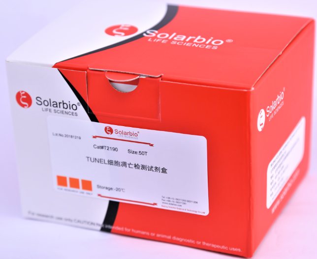
- T2190
- 北京
- 2025年07月16日

企业认证
相关产品推荐更多 >


万千商家帮你免费找货
0 人在求购买到急需产品
- 详细信息
- 询价记录
- 文献和实验
- 技术资料
- 库存:
大量现货
- 英文名:
TUNEL Apoptosis Assay Kit
- 保质期:
1年
- 供应商:
索莱宝
- 保存条件:
-20℃
- 规格:
50T
Ex/Em (nm) 556/579
Overview 
Printer Friendly Version
Ex/Em (nm)556/579MWN/ACAS #N/ASolventN/AStorageF/D/LCategoryCell Analysis
Cell Cytotoxicity
RelatedApoptosis and Cytotoxicity
Cell Apoptosis
Biochemical Assays
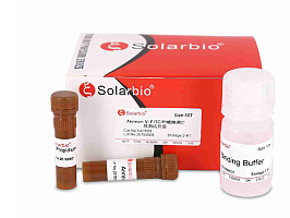
DNA fragmentation represents a characteristic of late stage apoptosis. DNA fragmentation in apoptotic cells can be detected by terminal deoxynucleotidyl transferase (TdT)-mediated dUTP nick end labeling (TUNEL). The TUNEL assay relies on the presence of nicks in the DNA which can be identified by TdT, an enzyme that catalyzes the addition of dUTPs that are secondarily labeled with a marker. All the existing TUNEL assays contain the highly toxic sodium cacodylate which might induces apoptosis and also decrease DNA production and DNA strands. Our Cell Meter™ TUNEL Apoptosis Assay Kit uses proprietary buffer system free of sodium cacodylate. The kit is based on incorporation of a fluorescence dye TF3 modified deoxyuridine 5'-triphosphates (TF3-dUTP) at the 3' OH ends of the DNA fragments that form during apoptosis. The assay is optimized for the direct detection of apoptosis in either detached or attached cells without using antibody. The kit provides all the essential components with an optimized assay protocol. It is suitable for fluorescence microplate reader, fluorescence microscope, or flow cytometer. Its signal can be easily detected at Ex/Em = 550nm/590 nm.
Quick Preview
This protocol only provides a guideline, and should be modified according to your specific needs.
Note: Thaw Components C at room temperature, keep Components A and B on ice before use.
1. Culture cells to an optimal density for apoptosis induction according to your specific protocol. We recommend about 30,000 to 50,000 cells/well for adherent cells grown in a 96-well microplate culture, or about 1 to 2 x 10⁶ cells/mL for non-adherent cells. At the same time, culture a non-induced negative control cell population at the same density as the induced population for every labeling condition. Here are a few examples for inducing apoptosis in suspension culture:
1) Treat Jurkat cells with 2 μg/ml camptothecin for 3 hours.
2) Treat Jurkat cells with 1 μM staurosporine for 3 hours.
3) Treat HL-60 cells with 4 μg/ml camptothecin for 4 hours.
4) Treat HL-60 cells with 1 μM staurosporine for 4 hours.
2. Fixation and Permeabilization
2.1 Remove cell media.
2.2 Add 100 μL/well/96-well plate of 4% formaldehyde fixative buffer (not supplied) to each well.
Note: For non-adherent cells, add desired amount (such as 2 x 10⁶ cells/mL) of 4% formaldehyde fixative buffer.
2.3 Incubate plates for 20 to 30 minutes at room temperature.
2.4 Remove fixative.
Optional: add 100 μL/well/96-well plate of the permeabilization reagent (0.2% Triton X-100 in PBS, not supplied) after the fixation if needed, and incubate the plate for 10 minutes at room temperature.
2.5 Wash the cells with PBS 2-3 times.
Optional: You may also prepare a positive control for TUNEL reaction using DNAase I by digesting cells with DNAase I for 30 min at room temperature before proceed to TUNEL reaction (Step 3)
相关产品
CA1030 Annexin V PE/7-AAD 凋亡检测试剂盒
CA1040 AnnexinV Alexa Fluor488/PI 凋亡检测试剂盒
CA1050 AnnexinV Alexa Fluor647/PI 凋亡检测试剂盒
BC3810 Caspase-1活性测定试剂盒
C6700 CCCP 细胞凋亡诱导剂(50mM)
CA1020 ANNEXIN V- FITC/PI 细胞凋亡检测试剂盒
G3680 细胞凋亡-Hoechst染色试剂盒
CA1120 细胞凋亡-Hoechst染色试剂盒
参考文献:
《The establishment of clonally derived chicken embryonic fibroblast cell line (CSC) with high transfection efficiency and ability as a feeder cell》 作者:Ruifeng Zhao , Jing Jin , Xinyu Sun , Kai Jin , Man Wang , Mahmoud F. Ahmed , Qisheng Zuo , Yani Zhang ,Zhenhua Zhao , Guohong Chen , Bichun Li 期刊:Journal of cellular Biochemistry 影响因子:3.062 PMID:30076744
《Crizotinib induces apoptosis of lung cancer cells through JAK-STAT pathway》 作者:Hongmin Lu Shibo Wu Huafei Chen Ying Huang Guoqin Qiu Lingxiang Liu Yong Li 期刊:Oncology Letters 影响因子:1.871 PMID:30333870
《Knockdown of TLR4 attenuates high glucose-induced podocyte injury via the NALP3/ASC/Caspase-1 signaling pathway》 作者:Yang Liu,Zhonggao Xu,Fuzhe Ma,Ye Jia,Guannan Wang 期刊:Biomedicine & Pharmacotherapy 影响因子:3.743 PMID:30257355
风险提示:丁香通仅作为第三方平台,为商家信息发布提供平台空间。用户咨询产品时请注意保护个人信息及财产安全,合理判断,谨慎选购商品,商家和用户对交易行为负责。对于医疗器械类产品,请先查证核实企业经营资质和医疗器械产品注册证情况。
- 作者
- 内容
- 询问日期
 文献和实验
文献和实验的细胞几乎没有DNA的断裂,因而没有3'-OH形成,很少能够被染色。由此,TUNEL成为了检测DNA片段化(细胞凋亡)的最常用方法。(以TUNEL法细胞凋亡检测试剂盒(增强型,绿色荧光)为例) TUNEL检测:使用20 U/ml Dnase处理Hela细胞10分钟。 二、TUNEL实验中关键步骤 1. 充分脱蜡和水化。脱蜡前可先将切片在 60ºC烤片 20 min,再使用二甲苯脱蜡2次,每次 5-10 min;而水化一般建议用梯度乙醇从高浓度到低浓度浸洗,便于后期试剂能充分、均匀的结合反应
用了许多方法检测小鼠的肝脏、脑部、睾丸等组织的细胞凋亡,其中 TUNEL 法做的最多,我购买的试剂盒主要是 roche、 promega 公司,结果大多数成功,偶尔也有失败,下面我把实验中的关键问题与大家共享一下,同时也希望能对初学者有所帮忙。 TUNEL 法的实验原理 基本原理:对不同组织切片先增加细胞膜通透性,然后让 rTDT 和生物标记的 dTUTP 进入细胞内,在 rTDT 的辅助下 dTUTP 与核断裂的 DNA 3』-OH 结合,再用 HRP 标记的链霉
Annexin V是一种钙离子依赖性磷脂结合蛋白,与磷脂酰丝氨酸PS有高度亲和力,可通过细胞外侧暴露的PS与凋亡早期细胞胞膜结合,将Annexin V标记上荧光染料利用流式细胞仪或荧光显微镜可检测细胞凋亡。 由于凋亡晚期或坏死细胞膜完整性丧失,细胞核染色液可进入细胞内染色细胞核,搭配Annexin V使用可将处于不同凋亡时期的细胞区分开。 A.悬浮细胞: 按照实验方案进行凋亡诱导,300 g离心5 min,弃上清,收集细胞,用PBS洗涤细胞一次,轻轻重悬浮细胞并计数。 取1~5 ×
 技术资料
技术资料
