


- ¥1500
- ATCC、DSMZ、ECACC、RIKEN
- 江苏
- CL1310
- 2026年02月02日

企业认证
相关产品推荐更多 >


万千商家帮你免费找货
0 人在求购买到急需产品
- 详细信息
- 询价记录
- 文献和实验
- 技术资料
- 英文名:
MCF 10A
- 库存:
100万
- 供应商:
欣润生物
- 肿瘤类型:
是
- 细胞类型:
细胞系
- ATCC Number:
CL1310
- 品系:
人
- 组织来源:
乳腺
- 相关疾病:
无
- 物种来源:
人源
- 免疫类型:
不详
- 细胞形态:
上皮型
- 是否是肿瘤细胞:
否
- 器官来源:
乳腺
- 运输方式:
新鲜或干冰
- 年限:
成年
- 生长状态:
贴壁生长
- 规格:
T25方瓶
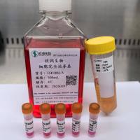
- 细胞名称:MCF 10A细胞(人正常乳腺上皮细胞)
- 形态:上皮型,贴壁生长
- 含量:>1x10⁶ 个/瓶
- 污染:支原体、细菌、酵母和真菌检测为阴性
- 规格:T25瓶或者1mL冻存管包装
二、细胞接收后的处理:
1、贴壁细胞
- 收到T25方瓶细胞后,请检查是否漏液,如果漏液,请拍照片发给我们(冻存管细胞收到后直接37℃水浴复苏或直接放置于液氮中长期储存)。
- 请先在显微镜下确认细胞生长状态,去掉封口膜并将T25瓶置于37℃培养约2-3h。
- 弃去T25瓶中的培养基,换用新鲜的完全培养基。
- 如果细胞长满(90%以上)请及时进行细胞传代。
- 接到细胞次日,请检查细胞是否污染,若发现污染或疑似污染,请及时与我们取得联系。
2、悬浮细胞
- 收到细胞后,请检查是否漏液,如果漏液,请拍照片发给我们。
- 请先在显微镜下确认细胞生长状态,去掉封口膜并将15ml离心管置于37℃培养约2-3h。
- 1200rpm离心5min,弃去15ml离心管中的培养基,细胞沉淀用新鲜的完全培养基重悬并培养。
- 如果细胞长满(90%以上)请及时进行细胞传代。
- 接到细胞次日,请检查细胞是否污染,若发现污染或疑似污染,请及时与我们取得联系。
本公司的细胞培养操作规程,供参考
一、培养基及培养冻存条件准备:
- 准备DMEM/F12培养基,90%;优质胎牛血清,10%。
- 培养条件: 气相:空气,95%;二氧化碳,5%。 温度:37℃,培养箱湿度为70%-80%。
- 冻存液:90%血清,10%DMSO,现用现配。液氮储存。
对于贴壁细胞,传代可参考以下方法:
- 弃去培养上清,用不含钙、镁离子的PBS润洗细胞1-2次。
- 加2ml消化液(0.25%Trypsin-0.53mM EDTA)于培养瓶中,置于37℃培养箱中消化2-3分钟,然后在显微镜下观察细胞消化情况,若细胞大部分变圆并脱落,迅速拿回操作台,轻敲几下培养瓶后加入3ml此细胞的培养基终止消化。
- 轻轻吹打后吸出,移入15ml离心管中,在1200RPM条件下离心5分钟,弃去上清液,加入1mL培养液后吹匀。
- 移入到事先准备好的含有5ml培养基的T-25培养瓶中或含有14ml培养基的T-75培养瓶中培养。
3)细胞冻存:待细胞生长状态良好时,可进行细胞冻存。贴壁细胞冻存时,先要消化处理并进行细胞计数。消化方法按照细胞传代方法的1-3步骤进行,最后的重悬液使用血清。悬浮细胞直接计数后离心,用血清重悬浮,加DMSO至最终浓度为10%。加入DMSO后迅速混匀,按每1ml的数量分配到冻存管中。本公司按每个冻存管细胞数目大于1X10⁶个细胞冻存。
注意事项:
1. 收到冻存管细胞后,若发现干冰已挥发干净、冻存管瓶盖脱落、破损及细胞有污染,请立即与我们联系。
2. 所有动物细胞均视为有潜在的生物危害性,必须在二级生物安全台内操作,并请注意防护,所有废液及接触过此细胞的器皿需要灭菌后方能丢弃。
3. 细胞用途:仅供科研使用。
发货方式:
复苏后发货:我们复苏细胞后发货,货期一周左右,免运费。(气温较好建议复苏后发货)
冻存发货(干冰运输):需额外增加干冰运费,选择干冰运输的我们发两管细胞,为了保证客户接种可靠性多发一管。(气温低于0℃须冻存发货)
细胞发货采取专业的运输包装,并选择最快捷的运输方式(顺丰速运或其他空运快递)
Isolation and Characterization of a Spontaneously Immortalized Human Breast Epithelial Cell Line, MCF-10
Two sublines of a breast epithelial cell culture, MCF-10, derived from human fibrocystic mammary tissue exhibit immortality after extended cultivation in low calcium concentrations (0.03-0.06 mM) and floating transfers in low calcium (MCF-10F), or by trypsin-Versene passages in the customary (normal) calcium levels, 1.05 mM (MCF-10A). Both sublines have been maintained as separate entities after 2.3 years (849 days) in vitro and at present have been in culture for longer than 4 years. MCF-10 has the characteristics of normal breast epithelium by the following criteria: (a) lack of tumorigenicity in nude mice; (b) three-dimensional growth in collagen; (c) growth in culture that is controlled by hormones and growth factors; (d) lack of anchorage-independent growth; and (e) dome formation in confluent cultures. Cytogenetic analysis prior to immortalization showed normal diploid cells; although later passages showed minimal rearrangement and near-diploidy, the immortal cells were not karyotypically normal. The emergence of an immortal culture in normal calcium media was not an inherent characteristic of the original tissue from which MCF-10 was derived since reactivated cryo-preserved cells from cultures grown for 0.3 and 1.2 years in low calcium were incapable of sustained growth in normal calcium.
Delineating Genetic Alterations for Tumor Progression in the MCF10A Series of Breast Cancer Cell Lines
To gain insight into the role of genomic alterations in breast cancer progression, we conducted a comprehensive genetic characterization of a series of four cell lines derived from MCF10A. MCF10A is an immortalized mammary epithelial cell line (MEC); MCF10AT is a premalignant cell line generated from MCF10A by transformation with an activated HRAS gene; MCF10CA1h and MCF10CA1a, both derived from MCF10AT xenografts, form well-differentiated and poorly-differentiated malignant tumors in the xenograft models, respectively. We analyzed DNA copy number variation using the Affymetrix 500 K SNP arrays with the goal of identifying gene-specific amplification and deletion events. In addition to a previously noted deletion in the CDKN2A locus, our studies identified MYC amplification in all four cell lines. Additionally, we found intragenic deletions in several genes, including LRP1B in MCF10CA1h and MCF10CA1a, FHIT and CDH13 in MCF10CA1h, and RUNX1 in MCF10CA1a. We confirmed the deletion of RUNX1 in MCF10CA1a by DNA and RNA analyses, as well as the absence of the RUNX1 protein in that cell line. Furthermore, we found that RUNX1 expression was reduced in high-grade primary breast tumors compared to low/mid-grade tumors. Mutational analysis identified an activating PIK3CA mutation, H1047R, in MCF10CA1h and MCF10CA1a, which correlates with an increase of AKT1 phosphorylation at Ser473 and Thr308. Furthermore, we showed increased expression levels for genes located in the genomic regions with copy number gain. Thus, our genetic analyses have uncovered sequential molecular events that delineate breast tumor progression. These events include CDKN2A deletion and MYC amplification in immortalization, HRAS activation in transformation, PIK3CA activation in the formation of malignant tumors, and RUNX1 deletion associated with poorly-differentiated malignant tumors.
风险提示:丁香通仅作为第三方平台,为商家信息发布提供平台空间。用户咨询产品时请注意保护个人信息及财产安全,合理判断,谨慎选购商品,商家和用户对交易行为负责。对于医疗器械类产品,请先查证核实企业经营资质和医疗器械产品注册证情况。
- 作者
- 内容
- 询问日期
 文献和实验
文献和实验Chicken intestinal epithelial cells were obtained from NEWGAINBIO company. Cells were cultured on 37℃, with 5% CO2, in the Ham’s F-12 Nutrient (DMEM/12) that contained the following supplementations: fetal bovine serum (5%), in-sulin (5 µg/mL), transferrin (5 µg/mL), selenium (5 ng/mL), epidermal growth factor (5 ng/mL) and penicillin-streptomycin (100–100 U/mL) for cell culturing (full DMEM/12). Experiments were performed with chicken intestinal epithelial cells and working solutions were prepared with plain DMEM/12 without supplementation. For the investigations, cells were seeded onto 96-well, 24-well or 6-well polystyrene cell culture plates.
Primary hVICs (passage 2) were cultured to 50–60% confluence and infected with pGMLV-SV40T-puro lentivirus (NewgainBio, Wuxi, China) at a multiplicity of infection of 80 supplemented with 5 µg/mL polybrene (Sigma-Aldrich, Buchs, Switzerland).
Tissue was cultured until cells became visible around the tissue, and when the fusion reached 90% (FIGURE 1A) §ask ¦lled with the prepared culturing medium was sent to the company for further immortalisation. Cell immortalisation was done for cell stability and longer-term use. Immortalised cells were cultured with 10% FBS and 1% PS in the DMEM medium. After the cells multiplied and merged, they were routinely passed and grown ( NEWGAINBIO Inc. Wuxi, Jiangsu, China) (FIGURE 1B-C).
Mouse primary cultured renal vascular ECs and VSMCs were obtained from Newgainbio company, which were tested by Factor VIII and α-smooth muscle actin (α-SMA), the marker of ECs and VSMCs. RNeasy Mini Kit was used for RNA extraction, and the above protocols were repeated.
Porcine primary colon epithelial cells (Newgainbio company, Wuxi,China) were cultured in Dulbecco's Modified Eagle's Medium (Solarbio, Beijing, China) containing 10 % fetal bovine serum (BioInd, Kiryat shmona, Lsrael) at 37 ◦C and 5 % CO2 humidity.
我正在培养MCF-7细胞,在此介绍一下我给细胞传代的经验:细胞放在显微镜下观察,细胞无污染、退缩,等其长满培养瓶约70%-80%时即可进行传代操作,具体步骤如下:1、 打开瓶盖,弃上清,2.D-HANK‘S夜(以50ml培养瓶为例,下同)2、吸管清洗后弃之;3、加0.25%胰酶2吸管;4、轻摇晃后置入37度温箱;5、3-5分钟后置显微镜下观察,见细胞胞质退缩,变圆;6、吸去胰酶,加含10%FCS的RPMI1640 3吸管;7、用洗管吹打,见细胞脱落,培养液变混,即可吸出加到新的培养瓶中。注意
wrwy 因实验急需MCF7,SKBR3乳腺癌细胞,哪位好心人能交换,本实验有T47D,231.293等细胞。谢谢。 msniu 我有MCF-7,站内联系 chenrongjun2011 本人在广州做实验,需要用到SKBR3,能否相赠一点,请加QQ1765730072或电话15915836004 本文由丁香园论坛提供,想了解更多有用的、有意思的前沿资讯以及酷炫的实验
0:00 内容简介0 0:15 实验过程15 17:48 结论1068 1191_capture_2.flv本视频来源于网络,如有异议请联系我们,我们将在5个工作日内作出处理。

