


- ¥3500 - 4500
- 欣润生物(NEWGAINBIO)
- 江苏无锡
- IH1013
- 2026年04月22日

企业认证
相关产品推荐更多 >


万千商家帮你免费找货
0 人在求购买到急需产品
- 详细信息
- 询价记录
- 文献和实验
- 技术资料
- 英文名:
Human Umbilical Vein Endothelial Cells
- 库存:
100万
- 供应商:
欣润生物
- 肿瘤类型:
否
- 细胞类型:
永生化
- ATCC Number:
无
- 品系:
人源
- 组织来源:
脐静脉
- 相关疾病:
无
- 物种来源:
人源
- 免疫类型:
不详
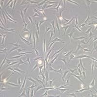
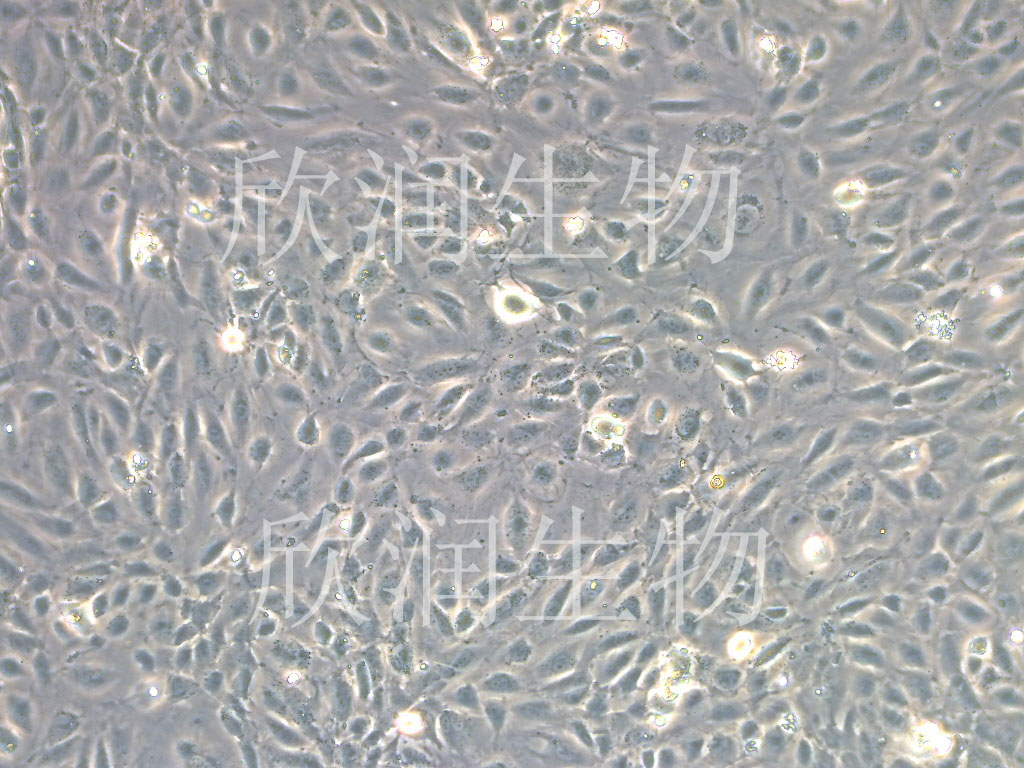
- 细胞形态:
梭形
- 是否是肿瘤细胞:
否
- 器官来源:
脐静脉
- 运输方式:
常温
- 年限:
/
- 生长状态:
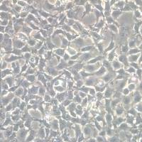
贴壁生长
- 规格:
T25方瓶
永生化人脐静脉内皮细胞简介:
产品描述:脐带是胎儿和胎盘之间的连系结构。形状如绳索,表面光滑透明,内含结缔组织和一支脐静脉,一对脐动脉。脐静脉内皮细胞来源于正常人脐静脉血管内膜表面,呈单层扁平分布。该细胞在合成和分泌细胞因子、维持血管内外凝血和纤溶的的动态平衡中起重要作用。内皮细胞还释放控制细胞增殖和调节血管壁紧张度的分子。这些过程都可以用培养的细胞进行体外研究,人脐静脉和动脉内皮细胞是这类研究中最常用的细胞类型。
产品货号:IH1013
产品类型: 永生化细胞
传代能力: 30代左右
产品形态: 梭形
培养基:永生化人脐静脉内皮细胞专用完全培养基,产品编号:IH1013-5
支原体:呈阴性
产品培养条件:37℃,5%CO2
发货方式:常温T25方瓶运输
货期:5-7左右货期
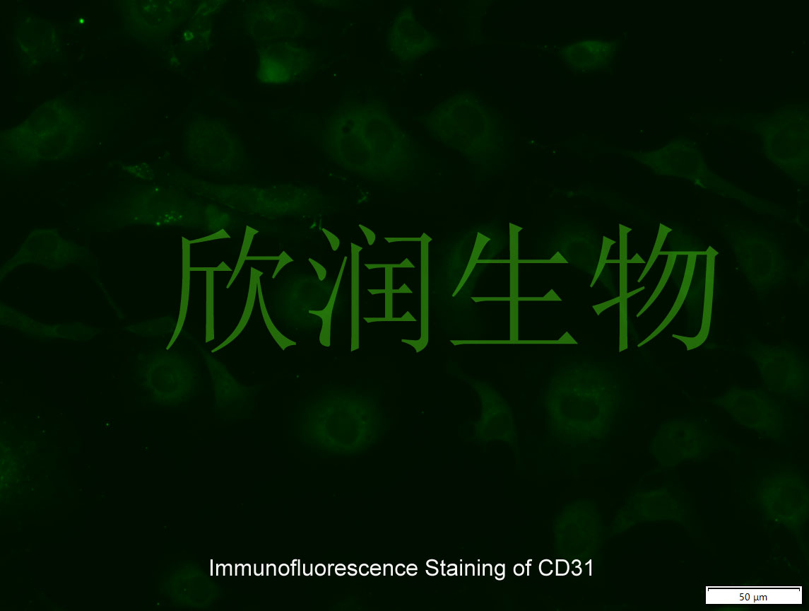
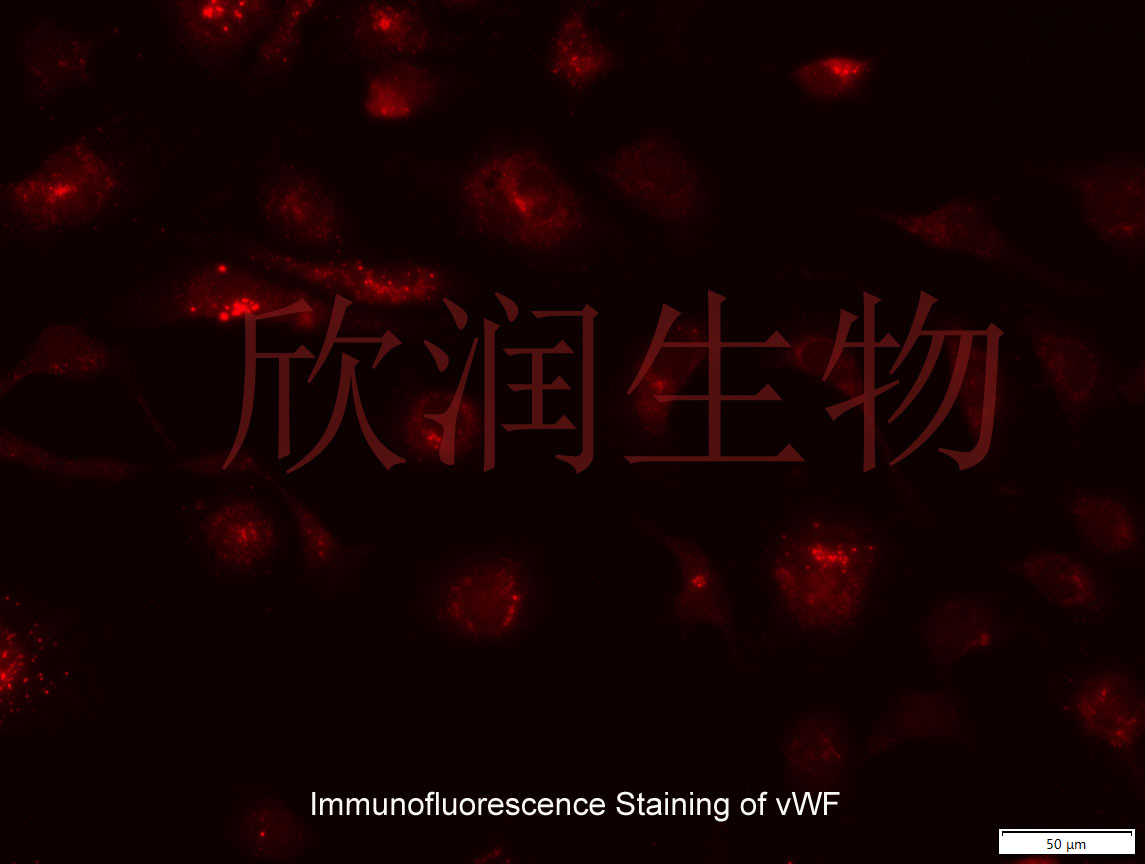
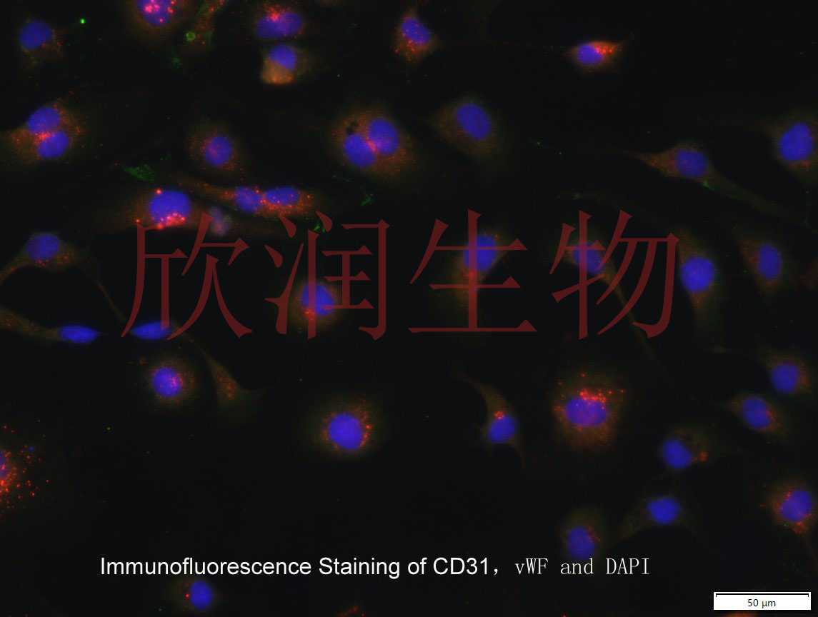
CD31和vWF抗体免疫荧光染色鉴定


Coenzyme Q10 prevents high glucose-induced oxidative stress in human umbilical vein endothelial cells
Hyperglycemia-induced oxidative stress plays a crucial role in the pathogenesis of vascular complications in diabetes. Although some clinical evidences suggest the use of an antioxidant reagent coenzyme Q 10 in diabetes with hypertension, the direct effect of coenzyme Q 10 on the endothelial functions has not been examined. In the present study, we therefore investigated the protective effect of coenzyme Q 10 against high glucose-induced oxidative stress in human umbilical vein endothelial cells (HUVEC). HUVEC exposed to high glucose (30mM) exhibited abnormal properties, including the morphological and biochemical features of apoptosis, overproduction of reactive oxygen species, activation of protein kinase Cβ2, and increase in endothelial nitric oxide synthase expression. Treatment with coenzyme Q 10 strongly inhibited these changes in HUVEC under high glucose condition. In addition, coenzyme Q 10 inhibited high glucose-induced cleavage of poly(ADP-ribose) polymerase, an endogenous caspase-3 substrate. These results suggest that coenzyme Q 10 prevents
风险提示:丁香通仅作为第三方平台,为商家信息发布提供平台空间。用户咨询产品时请注意保护个人信息及财产安全,合理判断,谨慎选购商品,商家和用户对交易行为负责。对于医疗器械类产品,请先查证核实企业经营资质和医疗器械产品注册证情况。
- 作者
- 内容
- 询问日期
 文献和实验
文献和实验Chicken intestinal epithelial cells were obtained from NEWGAINBIO company. Cells were cultured on 37℃, with 5% CO2, in the Ham’s F-12 Nutrient (DMEM/12) that contained the following supplementations: fetal bovine serum (5%), in-sulin (5 µg/mL), transferrin (5 µg/mL), selenium (5 ng/mL), epidermal growth factor (5 ng/mL) and penicillin-streptomycin (100–100 U/mL) for cell culturing (full DMEM/12). Experiments were performed with chicken intestinal epithelial cells and working solutions were prepared with plain DMEM/12 without supplementation. For the investigations, cells were seeded onto 96-well, 24-well or 6-well polystyrene cell culture plates.
Primary hVICs (passage 2) were cultured to 50–60% confluence and infected with pGMLV-SV40T-puro lentivirus (NewgainBio, Wuxi, China) at a multiplicity of infection of 80 supplemented with 5 µg/mL polybrene (Sigma-Aldrich, Buchs, Switzerland).
Tissue was cultured until cells became visible around the tissue, and when the fusion reached 90% (FIGURE 1A) §ask ¦lled with the prepared culturing medium was sent to the company for further immortalisation. Cell immortalisation was done for cell stability and longer-term use. Immortalised cells were cultured with 10% FBS and 1% PS in the DMEM medium. After the cells multiplied and merged, they were routinely passed and grown ( NEWGAINBIO Inc. Wuxi, Jiangsu, China) (FIGURE 1B-C).
Mouse primary cultured renal vascular ECs and VSMCs were obtained from Newgainbio company, which were tested by Factor VIII and α-smooth muscle actin (α-SMA), the marker of ECs and VSMCs. RNeasy Mini Kit was used for RNA extraction, and the above protocols were repeated.
Porcine primary colon epithelial cells (Newgainbio company, Wuxi,China) were cultured in Dulbecco's Modified Eagle's Medium (Solarbio, Beijing, China) containing 10 % fetal bovine serum (BioInd, Kiryat shmona, Lsrael) at 37 ◦C and 5 % CO2 humidity.
人脐静脉内皮细胞( HUVEC )培养 1 ). 将15-20cm长的新生儿脐带放入无菌的PBS溶液中储存。 (注:4℃下最多贮存24小时,室温下不超过6小时,否则废弃) 2 ). 用一个钝头的针头扎入脐带静脉管中,用无菌的PBS溶液冲洗3-5次,将污血冲洗干净为止。 3 ). 用手术钳夹紧脐带下端,加入15ml 的胶原酶(1mg/ml)室温下消化15-20分钟,并不时上下摇动脐带。 4 ). 消化完后,将下端手术钳松开
实验材料: 1. 婴儿脐带; 2. 不含Ca 2+ 和Mg 2+ 的1×PBS,添加200000IU/L青霉素、200mg/L链霉素,pH7.2; 3. 培养用液:M199培养液(含20%小牛血清);0.125%胰蛋白酶-0.01%EDTA(1:1,V:V)混合消化液;D-Hanks液、100IU/ml青霉素和100μg/ml链霉素; 4. 培养器具:玻璃插管与输液胶管,培养瓶或皿、白内障、眼科剪、镊子等; 培养方法: 1.
的二抗50μl(异硫氰酸标记羊抗鼠IgG,北京),放入37℃2h后,用PBS洗涤3次。然后立即在荧光显微镜下观察,摄片。 血管内皮细胞鉴定结果: VWF相关抗原间接免疫荧光检查,原代及传代培养的内皮细胞浆中有黄绿色的荧光着色,胞核呈黑绿色,整个细胞轮廓清楚。作为对照的内皮细胞片子中,偶见绿色斑点散发荧光,未见细胞轮廓。 5.内皮细胞体外生长情况 用胰蛋白酶消化的人脐静脉内皮细胞,接种在24孔塑料培养板各孔内4h后,大部分细胞贴壁。早期细胞呈小多角、球形、呈团状,少数细胞伸展,48~72h
 技术资料
技术资料
