

上皮间充质转化(EMT)ChIP qPCR芯片 Epithe
lial to Mesenchymal Transition (EMT) EpiTect ChIP qPCR Array- 询价
- 美国
- Epithelial to Mesenchymal Transition (EMT) EpiTect ChIP qPCR Array 上皮间充质转化(EMT)ChIP qPCR芯片
- 2026年02月02日

企业认证
相关产品推荐更多 >


万千商家帮你免费找货
0 人在求购买到急需产品
- 详细信息
- 询价记录
- 文献和实验
- 技术资料
- 服务名称:
上皮间充质转化(EMT)ChIP qPCR芯片
- 提供商:
SAbiosciences
技术服务网址:http://www.yingbio.com/
服务热线:400-696-6643、 18019265738
邮箱:daihp@yingbio.com 、 huizhang1228@foxmail.com
Epithelial to Mesenchymal Transition (EMT) EpiTect ChIP qPCR Array
上皮间充质转化(EMT)ChIP qPCR芯片
| Product | Species | Technology | Cat. No. |
| Epithelial to Mesenchymal Transition (EMT) EpiTect ChIP qPCR Array | Human | Histone Modifications | GH-090A |
| Epithelial to Mesenchymal Transition (EMT) EpiTect ChIP qPCR Array | Mouse | Histone Modifications | GM-090A |
The EpiTect Chip qPCR Arrays are intended for molecular biology applications. This product is not intended for the diagnosis, prevention, or treatment of a disease.
上皮间充质转化(EMT)ChIP qPCR芯片用于研究上皮间充质转变(EMT)的84个关键基因的组蛋白修饰状态或“组蛋白密码”。组蛋白修饰调节染色质结构,从而对基因表达的表观遗传调控发挥关键作用。EMT和相互间质上皮过渡(MET)是参与肿瘤的转移和干细胞分化和发育的关键步骤。在EMT期间,上皮细胞失去他们的顶点性和基底极性,破坏其细胞间的紧密连接,降解基底膜细胞外基质成分成为可迁移的间质细胞。因此该芯片包括细胞表面受体,细胞外基质,细胞骨架基因介导的细胞黏附、迁移、运动和形态,控制细胞分化、发育、生长和增殖的基因,以及导致EMT和所有相关的进程的信号转导和转录因子基因。结果可助于进一步洞察组蛋白修饰在调节EMT依赖性肿瘤转移和干细胞分化和发展中的作用。利用这个芯片通过染色质免疫沉淀和实时定量PCR,可以很简易、可靠地分析组蛋白的化学修饰模式与EMT相关重要基因的关联。
EpiTect ChIP qPCR仅用于分子生物学应用。本产品不用于疾病的诊断、预防和治疗。
Genes Up-Regulated During EMT: AHNAK, BMP1, CALD1, CDH2 (N-cadherin), COL1A2, COL3A1, COL5A2, FN1, FOXC2, GNG11, GSC, IGFBP4, ITGA5, ITGAV, MMP2 (Gelatinase A), MMP3, MMP9 (Gelatinase B), MSN, SERPINE1 (PAI-1), SNAI1, SNAI2, SNAI3, SOX10, SPARC, STEAP1, TCF4, TIMP1, TMEFF1, TMEM132A, TWIST1, VCAN, VIM, VPS13A, WNT5A, WNT5B.
Genes Down-Regulated During EMT: CAV2, CDH1 (E-cadherin), DSP, FGFBP1, IL1RN, KRT19, MITF, MST1R, NUDT13, PPPDE2, RGS2, SPP1 (Osteopontin), TFPI2, TSPAN13.
Genes with Known Histone Modifications during EMT:
Increased H3K4me3: AHNAK, AKT1, BMP1, CALD1, CAV2, CDH2 (N-cadherin), CTNNB1, FN1, FZD7, GNG11, GSK3B, IGFBP4, ILK, ITGA5, MAP1B, MITF, MMP2 (Gelatinase A), RGS2, SERPINE1 (PAI-1), SNAI1, SNAI2, SPARC, TCF4, TGFB1, TGFB2, TGFB3, TIMP1, TMEFF1, TSPAN13, VIM, VPS13A, WNT5A.
Decreased H3K27me3: DSP, FGFBP1, GSC, IL1RN.
Differentiation & Development: AKT1, BMP1, BMP2, BMP7, COL3A1, COL5A2, CTNNB1, DSP, ERBB3, F11R, FOXC2, FZD7, GSC, KRT14, MITF, MST1R, NODAL, NOTCH1, PTP4A1, SMAD2, SNAI1, SNAI2, SOX10, TGFB2, TGFB3, TMEFF1, TWIST1, VCAN, WNT11, WNT5A, WNT5B.
Morphogenesis: CTNNB1, FOXC2, PPP3R1, RAC1, SMAD2, SNAI1, SOX10, TGFB1, TGFB2, TGFB3, TWIST1, WNT11, WNT5A.
Cell Growth & Proliferation: AKT1, BMP1, BMP2, BMP7, CAV2, CTNNB1, EGFR, ERBB3, FGFBP1, FOXC2, HIF1A, IGFBP4, ILK, MST1R, NODAL, PDGFRB, TGFB1, TGFB2, TGFB3, TIMP1, VCAN.
Migration & Motility: CALD1, CAV2, EGFR, FN1, ITGB1, MSN, MST1R, NODAL, PDGFRB, RAC1, STAT3, TGFB1, VIM.
Cytoskeleton: CAV2, KRT7, MAP1B, PLEK2, RAC1, VIM.
Extracellular Matrix & Cell Adhesion: BMP1, BMP2, BMP7, CDH1 (E-cadherin), CDH2 (N-cadherin), COL1A2, COL3A1, COL5A2, CTGF, CTNNB1, DSC2, EGFR, ERBB3, F11R, FN1, FOXC2, ILK, ITGA5, ITGAV, ITGB1, MMP2 (Gelatinase A), MMP3, MMP9 (Gelatinase B), PTK2, RAC1, SERPINE1 (PAI-1), SPP1 (Osteopontin), TGFB1, TGFB2, TIMP1, VCAN.
Signaling Pathways:
Estrogen Receptor: CAV2, ESR1 (ERa), KRT19, TGFB3.
G-Protein Coupled Receptor: AKT1, FZD7, GNG11, RAC1, RGS2.
Integrin-Mediated: COL3A1, CTGF, ILK, ITGA5, ITGAV, ITGB1, PTK2.
Notch: FOXC2, NOTCH1.
Receptor Tyrosine Kinase: EGFR, ERBB3, PDGFRB, RGS2, SPARC.
TGFb / BMP: BMP1, BMP2, BMP7, COL3A1, SMAD2, SMAD4, TGFB1, TGFB2, TGFB3.
WNT: CTNNB1, FZD7, GSK3B, WNT11, WNT5A, WNT5B.
Transcription Factors: CTNNB1, ESR1 (ERa), FOXC2, GSC, MITF, NOTCH1, SIP1, SMAD2, SNAI2, SNAI3, SOX10, STAT3, TCF4, TWIST1, ZEB2
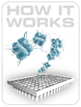
How it Works
The ChIP PCR array is a set of optimized real-time PCR primer assays on 96-well or 384-well plates for pathway or disease focused analysis of in vivo protein-DNA interactions. The ChIP PCR array performs ChIP DNA analysis with real-time PCR sensitivity and the multi-genomic loci profiling capability of a ChIP-on-chip. Simply mix your ChIP DNA samples with the appropriate ready-to-use PCR master mix, aliquot equal volumes to each well of the same plate, and then run the real-time PCR cycling program. (Download user manual)

What ChIP PCR Array Offers?
- Function or Disease Focused: Arrays represent a panel of genomic regions relevant to a biological function or disease state.
- Reliable & Sensitive: Arrays can analyze multiple genomic regions simultaneously with Real-Time PCR precision and sensitivity.
- Easy to Use Data Analysis: Download an easy-to-use Excel-based data analysis template [here]. Data analysis is based on the ΔΔCt method with normalization of the specific antibody and control IgG raw data to input raw data.
Layout and Controls: The PCR Arrays are available in both 96- and 384-well plates and are used to monitor the expression of 84 genes related to a disease state or pathway plus five housekeeping genes. Controls are also included on each array for ChIP DNA quality controls and general PCR performance.
You can easily perform a ChIP PCR Array experiment in your own laboratory, or send your samples to us and take advantage of our PCR Array Services.
Performance
EpiTect Chip qPCR Arrays provide the high sensitivity, specificity and reproducibility using SYBR-based real-time PCR technology.
Sensitivity
Together with our easy and fast One-Day ChIP kit, ChIP-Grade Antibody Kits, one million cells per assay as starting material provides 100% effective call rates.
| Ct Range
|
Percent Distribution of Ct Values | ||
| Input | H3K4me3 | Control IgG | |
| <24 | 0% | 27% | 0% |
| 25-30 | 100% | 60% | 0% |
| 30-35 | 0% | 13% | 96% |
| Absent Calls | 0% | 0% | 4% |
Table 1. ChIP PCR Arrays Analyze the Enrichment of 84 Genomic Sites with as Little as One Million Cells. P19 mouse embryonic carcinoma cells were prepared for ChIP Assay using the EpiTect Chip One-Day Kit and anti-H3K4me3 Antibody Kit. One million cells were used as starting material for each ChIP Assay. The purified ChIP DNA samples were characterized using Mouse Stem Cell Transcription Factor ChIP PCR Array with 1/100th of the ChIP DNA as template in each well. The Real-Time PCR results demonstrate 100 % effective call rates for the Input Fraction (Ct < 30). The difference of Ct value between the anti-H3K4me3 antibody and the control IgG fractions indicates the specific enrichment of the antibody, whereas the high Ct value of the control IgG fraction indicates the low background of the assay.
Reproducibility
The complete ChIP PCR Array System demonstrates a high degree of reproducibility across technical replicates, lots, instruments, and different handling, insuring reliable detection of differences in genomic DNA enrichment among biological samples.

Figure 5. Consistent Performance within the Same Plate or across Different Plates. Sonicated chromatin from HeLa cells (20 µg) was immunoprecipitated with 2 µg of anti-H3ac antibody or control IgG for 2 hours using the EpiTect Chip One-Day Kit. The obtained ChIP DNA samples were characterized in triplicates with EpiTect Chip qPCR primers specific for the active genes (GAPDH, RPL30, ALDOA), inactive genes (MYOD1, SERPINA), repetitive sequence (SAT2, SATa), and an ORF-free region (IGX1A) either within the same array plate or among different array plates in order to evaluate the intra- and inter-plate consistency. The anti-H3ac antibody enriched genomic DNA at active gene promoter regions with a high signal-to-noise ratio and a low co-efficiency of variation (less than 2.02%), irrespective of the type of assay (intra or inter-plate)

Figure 6. Consistent Performance with Various Amount of DNA Samples, Instruments or Handling Conditions. All experiments were performed in triplicates. Cells from MCF-7 (1 million per sample) were subjected to ChIP assay with anti-RNA Polymerase II (Pol 2) antibody followed by qPCR analysis of the proximal promoter of GAPDH, and an ORF-free region (IGX1A). Researcher A & B performed the PCR assays either in 96-well plate or 384-well plate format, on a Stratagene MX 3005 or an ABI 7900 Real-Time PCR instrument respectively. The same ChIP DNA samples were used which were stored for extended periods of time as indicated. The results demonstrate high reproducibility of PCR performance across technical replicates, lots, instruments, and differential handling.
Specific and Accurate ChIP-qPCR Detection
One prerequisite for ChIP PCR Array technology is the uniform and high
PCR amplification efficiency allowing a reciprocal comparison of ChIP enrichment among all genomic loci analyzed. The unique combination of SABiosciences' proprietary ChIP-qPCR primer design algorithm, rigorous validation of every ChIP-qPCR primer assay, and high performance SYBR Green master mix guarantees superior performance of EpiTect Chip qPCR Arrays.
A:

B:

Figure 7. Uniform Amplification Efficiency and Specific PCR Detection. 96 ChIP-qPCR primers were randomly picked from our genome-wide primer pool and analyzed for their performance. (A) All assays exhibit an average amplification efficiency of 99% with a 104.5% confidence interval between 102.5-105.2%, the uniform high amplification efficiency ensures accurate analysis of multiple genomic loci simultaneously using ΔΔCt method. (B) Each ChIP-qPCR primer assay is experimentally validated using dissociation (melt) curve analysis and agarose gel verification. Each pair of primers on PCR Array produces a single specific product as indicated by a single Dissociation Curve peak at a melting temperature (Tm) greater than 75 ºC, and PCR product was further validated on agarose gel for a single product of the predicted size without secondary products such as primer dimers
Application Examples
EpiTect Chip qPCR Arrays provide streamlined approaches to 1) Study biology or disease-focused gene regulation through histone modification and transcriptional regulatory network; 2) Monitor the dynamics of chromatin structure in the screening of function-specific epigenetic patterns; 3) Validate ChIP-on-chip or ChIP-seq results. The EpiTect Chip qPCR Arrays are also powerful tools for studying the mechanism contributing to gene expression changes observed by RT² Profiler PCR Arrays.
Below are listed a few examples of application data generated by our R&D group. To see the research using ChIP PCR Arrays published by the scientific community, please see our Publication List:http://www.sabiosciences.com/support_publication.php
Stem Cell Research
Stem cell differentiation into specific tissues involves the complex yet coordinated action of many transcription factors regulating not only tissue-specific genes, but also genes essential for differentiation itself. Histone modifications at the promoters of transcription factors are key mechanism regulating their expression. We used EpiTect Chip qPCR Arrays and RT² PCR Arrays to monitor the dynamic coordination of epigenetic modification and gene expression during retinoic acid (RA) induced differentiation of P19 mouse embryonic carcinoma cells (Figure 1). This RA treatment differentiates pluripotent P19 cells into somatic cells (Figure 2). The EpiTect Chip qPCR Array data showed that both gene expression and histone modifications on key transcription factors were changed in a dynamic manner through the course of P19 cell differentiation (Figure 3).

Figure 1. Schematic Representation of Pluripotency-Associated Gene Dynamics throughout Stem Cell Differentiation

Figure 2. Retinoic Acid (RA) Differentiation of Mouse Embryonic Carcinoma P19 Cells.

Figure 3. Dynamic Epigenetic Alternations and Gene Expression Changes during RA-Induced P19 Differentiation. ChIP PCR Arrays and RT² PCR Arrays were used to monitor the changes in gene expression levels and histone modification marks (H3Ac, H3K4me3, H3K27me3, and H3K9me3). The promoter region and expression levels of 84 key stem cell transcription factors were simultaneously analyzed during RA-induced neurogenesis of P19 cells at various time points (day 0, 4, and 8). Primer sets for the +1kb region downstream of the transcription start sites of the 84 genes and 12 control regions were preloaded on the ChIP PCR Array. Cluster analysis (http://www.sabiosciences.com/chippcrarray_data_analysis.php) of histone marks and mRNA level changes for the 84 genes were visualized as a heat map to represent the fold-differences during the RA-induced differentiation at the specified time points.
Characterize the Pattern of Histone Modifications
EpiTect Chip qPCR Arrays can be used to monitoring differential histone modifications across a gene.

Figure 4. The Custom EpiTect Chip 30Kb Tiling Array Quickly Maps Histone Modifications Surrounding the Transcription Start Site (TSS) of CDKN1A Gene. EpiTect Chip Antibodies against modified histones (H3Ac, H3K4me2, H3K27me3), or NIS were used to precipitate chromatin from one million HeLa cells. Each ChIP DNA fraction was analyzed with Custom EpiTect Chip 30Kb Tiling Array representing 30 one-kb tile intervals across the promoter region of the CDKN1A gene. The results indicate the enrichment of histone markers for actively transcribed genes (H3Ac and H3K4me2) but not marks for transcriptional inactive genes (H3K27me3) in the genomic region surrounding the TSS of CDNK1A.
风险提示:丁香通仅作为第三方平台,为商家信息发布提供平台空间。用户咨询产品时请注意保护个人信息及财产安全,合理判断,谨慎选购商品,商家和用户对交易行为负责。对于医疗器械类产品,请先查证核实企业经营资质和医疗器械产品注册证情况。
- 作者
- 内容
- 询问日期
 文献和实验
文献和实验【讨论】有没有研究EMT的,请过来看一看(MMP 2/9对肾纤维化的正负作用!)!!
liyoukong 请问这里有没有人研究肾纤维化中上皮细胞间充质转分化的(Epithelial-mesenchymal transition,EMT),有些问题想跟大家深入讨论一下!(也许帖子放的板块不是很正确,不过如果放到临床版块去的话就很少有人会关注这样的基础研究,所以考虑再三还是把这个帖子放到这个版块来了,请见谅!) 首先我们知道肾小管上皮细胞EMT是肾纤维化发生发展的重要步骤和中心环节,阻断甚至逆转EMT可以非常有效的减轻纤维化程度,甚至达到治疗
最近小编读到一篇关于 m6A 修饰的文章,完全被震撼和折服了!!思路之清晰、手段之全面、机制之详尽,让小编甘愿奉上自己的膝盖……毫不夸张的说够得上 Cell 级别(但是为什么没发 Cell 呢?),其他生物学过程/领域完全或部分套用这篇文章的思路,10 +文章妥妥的!!强烈建议收藏!!长文预警!!研究背景和待解决的科学问题癌细胞的转移是癌症致死的最主要原因,超过 90% 的癌症死亡与转移有关。EMT(上皮细胞-间质细胞转化)则是癌细胞转移过程中极其重要的一个步骤:上皮细胞在变成间充质
RIP技术(RNA结合蛋白免疫沉淀),研究肿瘤发生和转移中microRNA失调的有力工具
分泌肿瘤具有抑制作用。 最新的研究发现,许多miRNA参与晚期癌症的特征--癌细胞扩散过程的调控,这些miRNA通过同时作用多条信号通路并靶向不同目标蛋白基因而对癌细胞扩散起激活或抑制的功能(详见下图)。 肿瘤转移中miRNA调控的信号通路。在癌症恶化过程中,通常表达上升的miRNA以红色方框表示,而表达下降的miRNA以灰色方框表示。有助于上皮细胞-间充质转化的蛋白编码基因以绿色方框表示。EMT:epithelial

