

- ¥1700 - 4000
- GeneTex

- 美国
- GTX109242
- 2025年07月12日
- WB, ICC/IF, IHC-P, IHC-Fr, FACS, IP, ELISA, Sandwich ELISA
- Rabbit
- Human, Mouse, Rat

企业认证
相关产品推荐更多 >

![Cyclin H antibody [AT3G6]](https://img1.dxycdn.com/2022/0328/743/6228661923760800453-14.jpg!wh200)
![Kir2.1 antibody [S112B-14]](https://img1.dxycdn.com/2022/0328/150/8998962973014400453-14.jpg!wh200)
![IL17A antibody [MT504] (Biotin)](https://img1.dxycdn.com/2022/0328/366/4010824969230800453-14.jpg!wh200)
万千商家帮你免费找货
0 人在求购买到急需产品
- 详细信息
- 询价记录
- 文献和实验
- 技术资料
- 免疫原:
Full length human Arginase 1 Recombinant protein.
- 亚型:
IgG
- 形态:
Liquid
- 保存条件:
Store as concentrated solution. Centrifuge briefly prior to opening vial. For short-term storage (1-2 weeks), store at 4ºC. For long-term storage, aliquot and store at -20ºC or below. Avoid multiple freeze-thaw cycles.
- 克隆性:
Polyclonal
- 标记物:
Unconjugated
- 适应物种:
Human, Mouse, Rat
- 保质期:
12 months from the shipping date of the product.
- 抗原来源:
Human
- 目录编号:
GTX109242
- 级别:
Primary Antibodies
- 库存:
Available
- 供应商:
GeneTex
- 宿主:
Rabbit
- 应用范围:
WB, ICC/IF, IHC-P, IHC-Fr, FACS, IP, ELISA, Sandwich ELISA
- 浓度:
0.53 mg/ml (Please refer to the vial label for the specific concentration.)
- 靶点:
Arginase 1
- 抗体英文名:
Arginase 1 antibody
- 抗体名:
Arginase 1 抗体
- 规格:
100 μl/25 μl
| 规格: | 100 μl | 产品价格: | ¥4000.0 |
|---|---|---|---|
| 规格: | 25 μl | 产品价格: | ¥1700.0 |

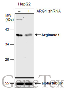
Non-transfected (–) and transfected (+) HepG2 whole cell extracts (30 μg) were separated by 10% SDS-PAGE, and the membrane was blotted with Arginase 1 antibody (GTX109242) diluted at 1:1000. The HRP-conjugated anti-rabbit IgG antibody (GTX213110-01) was used to detect the primary antibody.
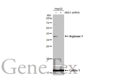
Non-transfected (–) and transfected (+) HepG2 whole cell extracts (30 μg) were separated by 12% SDS-PAGE, and the membrane was blotted with Arginase 1 antibody (GTX109242) diluted at 1:500. The HRP-conjugated anti-rabbit IgG antibody (GTX213110-01) was used to detect the primary antibody.
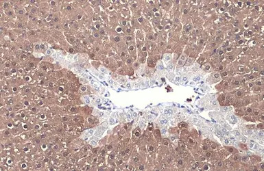
Arginase 1 antibody detects Arginase 1 protein at cytoplasm by immunohistochemical analysis.
Sample: Paraffin-embedded mouse liver.
Arginase 1 stained by Arginase 1 antibody (GTX109242) diluted at 1:500.
Antigen Retrieval: Citrate buffer, pH 6.0, 15 min
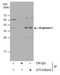
Immunoprecipitation of Arginase 1 protein from HepG2 whole cell extracts using 5 μg of Arginase 1 antibody (GTX109242).
Western blot analysis was performed using Arginase 1 antibody (GTX109242) diluted at 1:600.
EasyBlot anti-Rabbit IgG (GTX221666-01) was used as a secondary reagent.
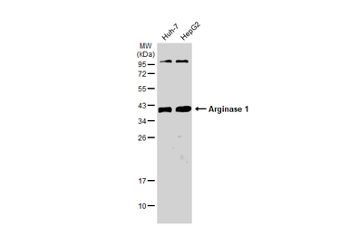
Various whole cell extracts (30 μg) were separated by 12% SDS-PAGE, and the membrane was blotted with Arginase 1 antibody (GTX109242) diluted at 1:1000. The HRP-conjugated anti-rabbit IgG antibody (GTX213110-01) was used to detect the primary antibody.
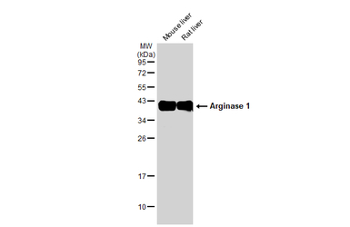
Various tissue extracts (50 μg) were separated by 12% SDS-PAGE, and the membrane was blotted with Arginase 1 antibody (GTX109242) diluted at 1:1000. The HRP-conjugated anti-rabbit IgG antibody (GTX213110-01) was used to detect the primary antibody.

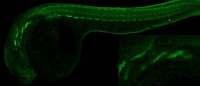
Arginase 1 antibody detects Arginase 1 protein by immunohistochemical analysis.
Sample: Frozen-sectioned mouse hippocampus.
Green: Arginase 1 stained by Arginase 1 antibody (GTX109242) diluted at 1:250.
Blue: Fluoroshield with DAPI (GTX30920).
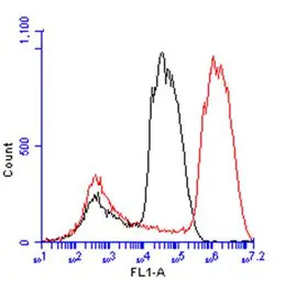
Arginase 1 antibody (GTX109242) detects ARG1 protein by flow cytometry analysis.
Sample: HepG2 cell.
Black: Unlabelled sample was used as a control.
Red: Arginase 1 antibody (GTX109242) dilution: 1:50.
Acquisition of 20,000 events were collected for FACS analysis.
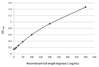
Sandwich ELISA detection of recombinant full-length Arginase 1 protein using GTX109242 as capture antibody at concentration of 5 μg/mL and GTX634218 as detection antibody at concentration of 1 μg/mL. Mouse IgG antibody (HRP) (GTX213111-01) was diluted at 1:10000 and used to detect the primary antibody.
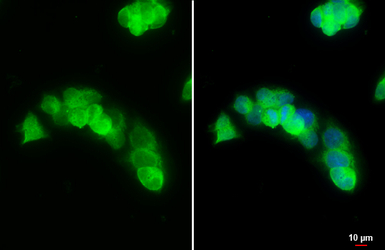
Arginase 1 antibody detects Arginase 1 protein at cytoplasm by immunofluorescent analysis.
Sample: HepG2 cells were fixed in 4% PFA at RT for 15 min.
Green: Arginase 1 stained by Arginase 1 antibody (GTX109242) diluted at 1:500.
Blue: Fluoroshield with DAPI (GTX30920).
Scale bar= 10 μm.
风险提示:丁香通仅作为第三方平台,为商家信息发布提供平台空间。用户咨询产品时请注意保护个人信息及财产安全,合理判断,谨慎选购商品,商家和用户对交易行为负责。对于医疗器械类产品,请先查证核实企业经营资质和医疗器械产品注册证情况。
- 作者
- 内容
- 询问日期
 文献和实验
文献和实验Vilhena ER et al., Behav Pharmacol 2021 (PMID:34320520)
Hoover AA et al., BMC Cancer 2020 (PMID:33028251)
Dichtl S et al., Sci Adv 2021 (PMID:34162546)
Tzeng HT et al., Cancers (Basel) 2021 (PMID:33809137)
Scherwitzl I et al., Mol Ther Oncolytics 2020 (PMID:32478167)
Sturgill ER et al., Oncoimmunology 2021 (PMID:33717655)
Nagumo Y et al., Microsurgery 2020 (PMID:33048377)
Lee YL et al., J Food Biochem 2020 (PMID:32964451)
Hanna A et al., Am J Physiol Heart Circ Physiol 2020 (PMID:32886000)
Di Domenico F et al., Redox Biol 2019 (PMID:30876754)
Zhang D et al., Nature 2019 (PMID:31645732)
Principe M et al., J Clin Endocrinol Metab 2020 (PMID:32785693)
Sui A et al., Invest Ophthalmol Vis Sci 2020 (PMID:32492108)
Wang W et al., Acta Biomater 2020 (PMID:32165192)
Arturo A Wilkins-Rodr?guez et al., Infect Immun 2020 (PMID:32312763)
Taisuke Okano et al., International Journal of Endocrinology 2020 2020()
Gualdr籀n-L籀pez M et al., Front Microbiol 2018 (PMID:29988527)
Pearse DD et al., Int J Mol Sci 2018 (PMID:30154346)
Polis B et al., Neurotherapeutics 2018 (PMID:30288668)
Glass EB et al., ACS Omega 2019 (PMID:31646220)
He Y et al., Int Immunopharmacol 2019 (PMID:31677497)
Tateyama H et al., Immunology 2019 (PMID:31520477)
Matsui S et al., Am J Pathol 2019 (PMID:31541644)
Barati S et al., J Cell Biochem 2019 (PMID:30963634)
Koizumi T et al., J Neuroinflammation 2019 (PMID:30971251)
Tziakas DN et al., Circulation 2019 (PMID:30717607)
Motterlini R et al., Redox Biol 2019 (PMID:30391826)
Carmona-Fontaine C et al., Proc Natl Acad Sci U S A 2017 (PMID:28246332)
Gomez D et al., Nat Med 2018 (PMID:30038218)
Yan G et al., Cancer Res 2018 (PMID:30012671)
Jang TJ et al., Pathol Res Pract 2018 (PMID:29970307)
Le Noci V et al., Oncoimmunology 2015 (PMID:26451303)
Shen P et al., BMC Microbiol 2017 (PMID:28835201)
Alabanza LM et al., J Immunol 2013 (PMID:23997223)
Zhu Y et al., Int J Mol Med 2017 (PMID:28627621)
Tavori H et al., J Lipid Res 2014 (PMID:25183802)
Pileri A et al., Virchows Arch 2017 (PMID:28321511)
Romano A et al., Oncotarget 2016 (PMID:27637084)
Wu J et al., J Neuroinflammation 2017 (PMID:28196545)
Royo F et al., Sci Rep 2017 (PMID:28211494)
Aalinkeel R et al., J Neuroimmune Pharmacol 2016 (PMID:28028734)
Mei S et al., PLoS One 2015 (PMID:26285119)
Ghosh M et al., J Neuroinflammation 2016 (PMID:26757726)
Harmon EY et al., J Am Heart Assoc 2014 (PMID:25516435)
Sward K et al., Physiol Rep 2013 (PMID:24303100)
Jindal A et al., Exp Mol Pathol 2015 (PMID:26112094)
Beachley VZ et al., Nat Methods 2015 (PMID:26480475)
Gladine C et al., Genes Nutr 2014 (PMID:25134659)
Yoshii A et al., Biol Reprod 2014 (PMID:24966392)
Sward K et al., PLoS One 2014 (PMID:24658465)
Guo Z et al., J Immunol 2013 (PMID:23514739)
Panebianco C et al., Cell Death Discov 2023 (PMID:37019893)
Anderson S et al., Biomedicines 2022 (PMID:36009546)
Huang J et al., Mol Metab 2023 (PMID:36481344)
Broome ST et al., J Mol Neurosci 2022 (PMID:35199308)
Jian Wang et al., Theranostics 2022 (PMID:35673563)
Le Liu et al., Front Pediatr 2022 (PMID:35733809)
Tingting Wang et al., Front Cell Dev Biol 2020 (PMID:33392187)
Libu?e Jansk? et al., Dis Model Mech 2021 (PMID:34407185)
Wang K et al., Transl Neurosci 2021 (PMID:34900345)
Ashenafi S et al., Am J Pathol 2022 (PMID:35092727)
Wu L et al., Theranostics 2022 (PMID:34976216)
Wilson AJ et al., Neoplasia 2022 (PMID:34933276)
Grzywa TM et al., Commun Biol 2021 (PMID:34893694)
Cai M et al., CNS Neurosci Ther 2021 (PMID:34370899)
上海西唐生物科技有限公司 021-55229872, 65333639 www.westang.com 人精氨酸酶 (Arginase)ELISA 试剂盒 ( 用于血清、血浆、细胞培养上清液和唾液等其它生物体液内 ) 原理 本实验采用双抗体夹心 ABC-ELISA 法。用抗人 Arginase 单抗包被于酶标板上,标准品和样品中的 Arginase与单抗结合,加入生物素化的抗人
Elabscience 巨噬细胞三大功能及检测方案全梳理,看完少走90%弯路!
悬液的表型鉴定和功能分析。巨噬细胞没有类似于T细胞(CD4/CD8)那样明确的单一区分抗原,因此需要使用一组标志物的组合来鉴定巨噬细胞。对于小鼠巨噬细胞,常用的组合包括F4/80、CD11b、Ly6C和THY-1等。通过多色荧光标记和流式细胞仪分析,可以在异质性细胞群中精确地鉴定和分选出巨噬细胞。 图5. C57BL/6J腹腔炎症小鼠取腹水样本,使用PE Anti-Mouse/Human/Monkey CD11b Antibody[M1/70](E-AB-F1081D)、Elab Fluor®


