

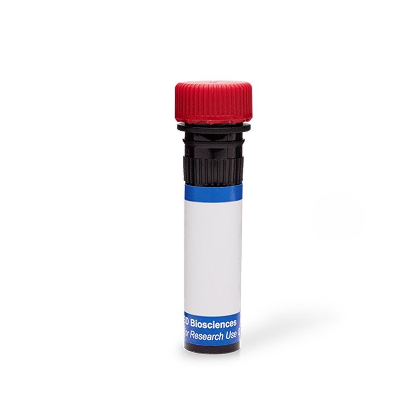
- ¥4337.0¥188特惠体验价
- BD
- 美国
- 566683
- 2026年10月23日
- Flow Cytometry (FCM)
- Mouse
- Human

企业认证
相关产品推荐更多 >

Rabbit anti-RelB Polyclonal Antibody【兔抗RelB多克隆抗体】- abs125092
¥2300
Rabbit anti-DIO3 Polyclonal Antibody【兔抗DIO3多克隆抗体】- abs145893
¥1500Rabbit anti-eIF4G Polyclonal Antibody【兔抗eIF4G多克隆抗体】- abs124822
¥2300
Rabbit anti-CYP2A6 Polyclonal Antibody【兔抗CYP2A6多克隆抗体】- abs153793
¥1600Mouse anti-PLIN3 Monoclonal Antibody【小鼠抗PLIN3单克隆抗体】- abs113586
¥1500
万千商家帮你免费找货
0 人在求购买到急需产品
相关阅读
源叶生物 十万种现货试剂速发,品质稳、品类全,实验不再等!Takara 秋促来袭,提取+反转录+定量套装仅 1200 元abinScience 体内级抗体 全年锁价 低至 ¥1200阿拉丁 22万+ 科研级产品现货速发,品质严控、科研无忧- 详细信息
- 文献和实验
- 技术资料
- 供应商:
BD Pharmingen
- 靶点:
CD3
- 级别:
RUO
- 克隆性:
单克隆
- 抗原来源:
Human
- 抗体英文名:
CD3 Antibody
- 抗体名:
CD3
- 标记物:
PE
- 宿主:
Mouse
- 适应物种:
Human
- 免疫原:
Sheep Erythrocyte Rosette-purified Human T Cells
- 亚型:
Mouse BALB/c x A/J, also known as CAF1 IgG2a, κ
- 形态:
液体
- 应用范围:
Flow Cytometry (FCM)
- 保存条件:
2-8℃避光保存
- 规格:
100 Tests
产品概述
Hu CD3 PE OKT3 100Tst 为 BD Pharmingen 品牌流式抗体,货号 566683,靶向 CD3,荧光标记 PE,克隆 OKT3 (RUO),规格 100 Tests。适用于 Flow cytometry (Routinely Tested) 检测,仅供科研使用(RUO)。
靶点与抗体说明
The OKT3 monoclonal antibody specifically recognizes the CD3 epsilon subunit (CD3e/CD3ε) of the CD3 complex which consists of four transmembrane proteins (γ, δ, ε, ζ) that are associated with the T cell antigen receptor (TCR) to form the CD3/TCR complex. The CD3 complex associates with either TCR αβ or TCR γδ heterodimers that are alternatively expressed by some thymocytes, T cells or NKT cells. The CD3 complex is required for the cell surface expression and signal-transducing functions of the TCR. The CD3 complex is expressed by ~60-85% thymocytes and by all peripheral mature T cells. CD3e is also known as T3E or TCRE. CD3e is a ~20 kDa unglycosylated type I transmembrane protein that is encoded by CD3E which belongs to the immunoglobulin superfamily (IgSF). CD3e has an Ig-like extracellular domain (ECD) and an immunoreceptor tyrosine-based activation motif (ITAM) in its cytoplasmic domain. The OKT3 antibody can reportedly fix complement, stimulate T cell proliferation and cytokine production, and block the binding of other human CD3e-specific antibodies including UCHT1 and SK7. R-Phycoerythrin (PE), is part of the BD family of Phycobiliprotein dyes. This fluorochrome is a multimeric fluorescent phycobiliprotein with excitation maximum (Ex Max) of 496 nm and 566 nm and an emissi
核心参数
| 货号 | 566683 |
| 品牌 | BD Pharmingen |
| 克隆号 | 克隆 OKT3 (RUO) |
| 宿主 | Mouse |
| 同种型 | Mouse BALB/c x A/J, also known as CAF1 IgG2a, κ |
| 荧光标记 | PE |
| 抗原 / 靶点 | CD3 |
| 反应种属 | Human |
| 验证应用 | Flow cytometry (Routinely Tested) |
| 推荐用量 | 5 µl |
| 免疫原 | Sheep Erythrocyte Rosette-purified Human T Cells |
| 规格 | 100 Tests |
| 存储缓冲液 | Aqueous buffered solution containing BSA and ≤0.09% sodium azide. |
荧光染料信息
PE R-Phycoerythrin (PE), is part of the BD family of Phycobiliprotein dyes. This fluorochrome is a multimeric fluorescent phycobiliprotein with excitation maximum (Ex Max) of 496 nm and 566 nm and an emission maximum (Em Max) at 576 nm. PE is designed to be excited by the Blue (488 nm), Green (532 nm) and Yellow-Green (561 nm) lasers and detected using an optical filter centered near 575 nm (e.g., a 575/26-nm bandpass filter). As PE is excited by multiple lasers, this can result in cross-laser excitation and fluorescence spillover on instruments with various combinations of Blue, Green, and Yellow-Green lasers. Please ensure that your instrument’s configurations (lasers and optical filters) are appropriate for this dye. 格式:PE最多激发源:496 nm, 566 nm最多发射源:576 nm
保存与运输
建议 2-8℃ 避光保存,请勿冷冻,避免反复冻融。 存储缓冲液:Aqueous buffered solution containing BSA and ≤0.09% sodium azide.
参考文献
Burns GF, Boyd AW, Beverley PC. Two monoclonal anti-human T lymphocyte antibodies have similar biologic effects and recognize the same cell surface antigen. J Immunol. 1982; 129(4):1451-1457. (Clone-specific: Blocking, Functional assay, Immunoprecipitation, Radioimmunoassay). Emmrich F. Selective stimulation of human CD4 and CD8 T-cells by crosslining the T-cell receptor with subset-specific differentiation antigens. In: McMichael AJ. A.J. McMichael .. et al., ed. Leucocyte typing III : white cell differentiation antigens. Oxford New York: Oxford University Press; 1987:203-206. Ernst DN, Shih CC. CD3 complex. J Biol Regul Homeost Agents. 2000; 14(3):226-229. (Biology). Horibe K, Knowles RW, Naito K, Morishima Y, Dupont B. Analysis of T lymphocyte antibody specificities: Comparison of serology with immunoprecipitation patterns. In: Bernard A. A. Bernard .. et al., ed. Leucocyte typing : human leucocyte differentiation antigens detected by monoclonal antibodies. Berlin New York: Springer-Verlag; 1984:212-224. Kung P, Goldstein G, Reinherz EL, Schlossman SF. Monoclonal antibodies defining distinctive human T cell surface antigens. Science. 1979; 206(4416):347-349. (Immunogen: Cytotoxicity, Flow cytometry, Radioimmunoassay). Kurrle R, Seyfert W, Trautwein A, Seiler FR. T cell activation by CD3 antibodies. In: Reinherz EL. Ellis L. Reinherz .. et al., ed. Leukocyte typing II. New York: Springer-Verlag; 1986:137-146. Li B, Wang H, Dai J, et al. Construction and characterization of a humanized anti-human CD3 monoclonal antibody 12F6 with effective immunoregulation functions. Immunology. 2005; 116(4):487-498. (Clone-specific: Blocking, Flow cytometry). Semnani R, Nutman TB, Corrado G, Hochman P, Shaw S, Van Seventer GA. Costimulation mediated by purified ICAM-1 and LFA-3 regulates differential stimulation and cytokine secretion of human "naive" and "memory" CD4+ T cells. In: Schlossman SF. Stuart F. Schlossman .. et al., ed. Leucocyte typing V : white cell differentiation antigens. Oxford: Oxford University Press; 1995:1488-1491. Touraine JL, Favrot MC, Ansary ME, Cordier G, de bouteiller O. Phenotype of prothymocytes from human bone marrow determined by monoclonal antibodies: Modification induced by thymic factots. In: Bernard A. A. Bernard .. et al., ed. Leucocyte typing : human leucocyte differentiation antigens detected by monoclonal antibodies. Berlin New York: Springer-Verlag; 1984:298-311. Tunnacliffe A, Olsson C, Traunecker A, Krissansen GW, Karjalainen
风险提示:丁香通仅作为第三方平台,为商家信息发布提供平台空间。用户咨询产品时请注意保护个人信息及财产安全,合理判断,谨慎选购商品,商家和用户对交易行为负责。对于医疗器械类产品,请先查证核实企业经营资质和医疗器械产品注册证情况。
 文献和实验
- 3) 及 CD45 -PE (HI30,白细胞标志) 抗体染色后,进行流式分析,死细胞以 DAPI 染色先行排除,左右分别为分选前后的细胞群分布。 总结: 几次实验的结果,使用 Fab-TACS® Gravity column 来从全血直接进行 CD3+ T 细胞分选,平均得率为 84.5%,纯度大于 90%,细胞存活率高于 95%,且目标细胞不含标记,贴近天然,使下游实验更精确。
Nature 综述导读:FDA 批准第 100 个抗体药物!抗体药物的研究方向在哪里?
Nature Reviews Drug Discovery 20, 10 (2021)单克隆抗体 (mAbs) 由 B 细胞产生,特异性靶向抗原。该产品能够得以大量生产制备归功于 Köhler 和 Milstein 于 1975 年发明的杂交瘤技术,该技术极大地提高了基础研究和临床应用的潜力,利用该技术,Schlossman 和他的合作者鉴定出一种针对特殊 T 细胞抗原的单克隆抗体 - OKT3,它针对的抗原便是 CD3。1981 年,他们在临床测试 OKT3 作为预防移植排斥的免疫抑制剂。1986 年
: 由此步骤开始,流出的溶液包含目标细胞,请使用新的试剂管收集!首先,加入 1 ml 的 biotin 洗脱液,孵育 2 min。其次,加入 10 ml biotin 洗脱液,洗脱目标细胞。d) 再加入 10 ml biotin 洗脱液,洗下剩余的目标细胞。最后两步骤所收集的液体含分选出的目标细胞。(4) 结果分析以下流式图为从 7.5 ml 全血中,选出 CD3+ T 细胞的数据。选后细胞以 CD3-APC(OKT-3) 及 CD45-PE(HI30,白细胞标志) 抗体染色后,进行流式分析,死细胞
 技术资料
暂无技术资料 索取技术资料