


- ¥3500 - 4500
- 欣润生物
- 江苏无锡
- IR9003-1
- 2025年12月13日

企业认证
相关产品推荐更多 >


万千商家帮你免费找货
0 人在求购买到急需产品
- 详细信息
- 文献和实验
- 技术资料
- 英文名:
Smooth muscle cells of rabbit aorta
- 库存:
10
- 供应商:
欣润生物
- 肿瘤类型:
NO
- 细胞类型:
永生化
- ATCC Number:
11222
- 品系:
新西兰兔
- 组织来源:
兔主动脉平滑肌
- 相关疾病:
无
- 物种来源:
兔
- 免疫类型:
不详
- 细胞形态:
梭形
- 是否是肿瘤细胞:
否
- 器官来源:
主动脉
- 运输方式:
常温
- 年限:
5年
- 生长状态:
贴壁生长
- 规格:
T25方瓶
永生化兔主动脉平滑肌细胞简介:
产品描述:平滑肌细胞对心血管疾病的发生起着极其重要的作用。心血管疾病的发生和发展的一个主要因素在于心血管平滑肌细胞转变成为了具有增殖能力的表型。近期的研究表明,平滑肌细胞能表达钙离子通道ICAM-1和VCAM-1,其中ICAM-1和VCAM-1的表达可能是造成血管壁炎症反应,并进一步造成心血管疾病的原因。因此,对血管平滑肌细胞的体外培养和研究可用来发现和确定新的血管疾病的靶向治疗方法。
产品货号:IR9003
产品类型: 永生化细胞
传代能力: 30代左右
产品形态: 成纤维细胞样
培养基:永生化兔主动脉平滑肌细胞完全培养基
支原体质控:呈阴性
产品培养条件:37℃,5%CO2
发货方式:T25瓶子常温发货
货期:1周左右货期
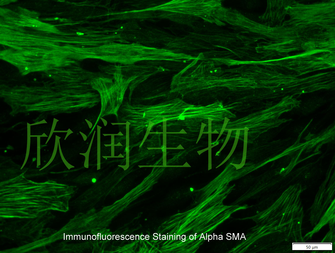
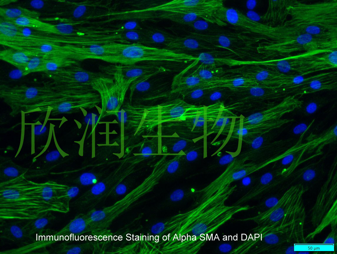
a-SMA免疫荧光染色


Contractility of smooth muscle cells of rabbit aorta in tissue culture
THE involvement of smooth muscle cells (SMC) from the media of elastic arteries has been implied in the aetiology of atherosclerosis 2 . Aortic tissue cultures have been studied repeatedly to examine the ability of SMC to synthetise macromolecular proteins and collagen and elastic fibres. Ross 3 has shown that aortic cultured cells retained all the morphological characteristics of SMC up to week 8 of culture. By week 4 microfibrils identified as elastic fibres appeared close to the cells. On the other hand, Ouzilou et al. 4 showed that the cultured aortic fragments can be used as a suitable model to study the metabolic properties of aortic smooth muscle. The explants retained, for at least a week, the ability to synthesise the proteins of the medial matrix at a rate that decreased with the age of the rabbits from which the aortic specimens were obtained.
风险提示:丁香通仅作为第三方平台,为商家信息发布提供平台空间。用户咨询产品时请注意保护个人信息及财产安全,合理判断,谨慎选购商品,商家和用户对交易行为负责。对于医疗器械类产品,请先查证核实企业经营资质和医疗器械产品注册证情况。
 文献和实验
文献和实验Chicken intestinal epithelial cells were obtained from NEWGAINBIO company. Cells were cultured on 37℃, with 5% CO2, in the Ham’s F-12 Nutrient (DMEM/12) that contained the following supplementations: fetal bovine serum (5%), in-sulin (5 µg/mL), transferrin (5 µg/mL), selenium (5 ng/mL), epidermal growth factor (5 ng/mL) and penicillin-streptomycin (100–100 U/mL) for cell culturing (full DMEM/12). Experiments were performed with chicken intestinal epithelial cells and working solutions were prepared with plain DMEM/12 without supplementation. For the investigations, cells were seeded onto 96-well, 24-well or 6-well polystyrene cell culture plates.
Primary hVICs (passage 2) were cultured to 50–60% confluence and infected with pGMLV-SV40T-puro lentivirus (NewgainBio, Wuxi, China) at a multiplicity of infection of 80 supplemented with 5 µg/mL polybrene (Sigma-Aldrich, Buchs, Switzerland).
Tissue was cultured until cells became visible around the tissue, and when the fusion reached 90% (FIGURE 1A) §ask ¦lled with the prepared culturing medium was sent to the company for further immortalisation. Cell immortalisation was done for cell stability and longer-term use. Immortalised cells were cultured with 10% FBS and 1% PS in the DMEM medium. After the cells multiplied and merged, they were routinely passed and grown ( NEWGAINBIO Inc. Wuxi, Jiangsu, China) (FIGURE 1B-C).
Mouse primary cultured renal vascular ECs and VSMCs were obtained from Newgainbio company, which were tested by Factor VIII and α-smooth muscle actin (α-SMA), the marker of ECs and VSMCs. RNeasy Mini Kit was used for RNA extraction, and the above protocols were repeated.
Porcine primary colon epithelial cells (Newgainbio company, Wuxi,China) were cultured in Dulbecco's Modified Eagle's Medium (Solarbio, Beijing, China) containing 10 % fetal bovine serum (BioInd, Kiryat shmona, Lsrael) at 37 ◦C and 5 % CO2 humidity.
一、摘要 目的:建立兔膀胱平滑肌细胞的分离、培养和鉴定的方法。方法:成年新西兰白兔两只(正常及梗阻各一只),采用酶法分离技术获得膀胱平滑肌细胞后于10%小牛血清的DMEM中培养,观察细胞形态和扩增情况,用爬片染色、电镜、蛋白质α-肌动蛋白(α-actin)鉴定细胞类型。结果:倒置显微镜下观察均呈“谷和峰”样结构、细胞爬片HE染色及电镜检查均证实为平滑肌细胞。免疫组化染色检测α-actin呈阳性反应。从细胞爬片HE染色和免疫组化染色检测α-actin呈阳性反应中我们发现该方法所的膀胱平滑肌细胞
实验材料: 1. 正常大兔主动脉 2. 不含Ca2+ 和Mg2+ 的1×PBS,添加200000IU/L青霉素、200mg/L链霉素,pH7.2 3. 消化液:0.125%胰蛋白酶,0.1%胶原酶Ⅰ 4. 细胞培养瓶(T25) 5. 手术刀、解剖剪、解剖镊、眼科剪,眼科镊 6. 网筛(100目) 7. 离心管(15ml、50ml) 实验方法: 1. 处死大兔,分离出主动脉,放入预冷的含双抗的1×PBS(pH=7.2)反复清洗3次。以洗去
粥样硬化的演变过程相似,已被广泛应用。 本实验通过腹主动脉球囊拉伤法,制备兔动脉粥样硬化模型。 腹主动脉球囊拉伤法,制备兔动脉粥样硬化模型 一、实验动物 新西兰兔,雄性,体重 2.5~3 kg。 二、实验材料 导管球囊:单腔动脉取血栓导管,Edwards,USA;高脂饲料。 三、实验方法 取体重为 2.5-3 kg 的新西兰兔,普通饲料适应性饲养 1 周,1 周后给予高脂饲料饲养并制备模型。 制备模型前空腹 12 小时,戊巴*比妥钠 (30 mg/kg) 经耳缘静脉麻醉动物, 穿刺
 技术资料
技术资料