互联网2013-11-13
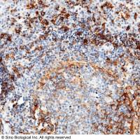
The following protocol used B6 mice that were infected with 200 embryonated Trichuris muris eggs and sacrificed 15 days post-infection. The tip of the cecum was removed, rinsed in 1X PBS pH 7.2, and fixed in 4% PFA. Following fixation, the tissue was embedded in paraffin and cut into 5 μM sections.
1. Deparaffinize and rehydrate the tissue section.
2. Perform heat-induced antigen retrieval by boiling the tissue section in 10mM pH 6.0 citrate buffer for 25 minutes.
3. Incubate the tissue section with blocking buffer for 20 minutes.
4. Incubate the tissue section overnight at 4?C with Rabbit Anti-Murine RELMα at 4.0 ng/mL in 1X PBS with 0.01% Triton-X and 0.5% BSA. Wash the slide twice for three minutes (1X PBS/0.05% Tween 20).
5. Incubate the tissue section with a fluorescent conjugated secondary antibody for 2 hours at room temperature. Wash the slide twice for three minutes.
6. Counterstain the tissue section with DAPI.
The following protocol used B6 mice lung sections that were injected with helminth Schistosoma mansoni eggs. Inflamed lung sections were fixed in 4% PFA. Following fixation, the tissue was embedded in paraffin and cut into 5 μM sections. Some tissue sections or cell preparations stained were from day 14 bleomycin-instilled mice.
4. Incubate the tissue section overnight at 4?C with Rabbit Anti-Murine RELMα at 0.2 μg/mL in 1X PBS with 0.01% Triton-X and 0.5% BSA. Wash the slide twice for three minutes (1X PBS/0.05% Tween 20).
相关产品推荐
IL4R 兔单抗(Anti-Human)
¥900
RELM beta Rabbit pAb(bs-5774R)-50ul/100ul/200ul
¥1180
CD14 Antibody, Rabbit MAb | CD14 兔单抗
¥800
UCF.ME 鼠源RNase抑制剂(Murine RNase inhibitor) GMP级别
¥1398
LYPD3 兔单抗(Anti-Human)
¥1100
相关问答
问
二抗anti-mouse可以代替anti-rat吗?
RABBIT ANTI HUMAN BETA AMYLOID (aa1-42)抗体做Elisa实验板的板包被,需要多少浓度的抗体
Anti-KAT5 / Tip60抗体
相关方法
🔥 免疫荧光(Immunofluorescence, IF)
2023-05-26
赤霉素对α-淀粉诱导合成的影响
2022-02-11
α 突触核蛋白操作指南
2023-08-08
推荐阅读
Detection of RTK Pathway Activation in Drosophila Using Anti-dpERK Immunofluorescence Staining
Teratology Studies in the Rabbit
Rabbit Whole Embryo Culture