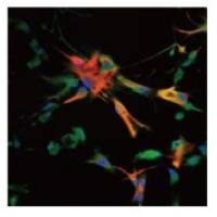
原代神经元培养
互联网
Protocol for the Primary Culture of Cortical and Hippocampal neurons
Solutions and media required:
Poly D-lysine/laminin solution - pdf
DM/KY - pdf
Optimem - pdf
Neuronal growth medium - pdf
Set-up for the dissection:
Day 1:
Add poly D-lysine/laminin solution to culture dishes/coverslips. Swirl the plate to ensure that the coating mix covers the entire bottom of the plate. Leave the dishes/coverslips in the 37oC/5% CO2 incubator overnight.
Day 2:
Wash the dishes/coverslips twice with sterile water; remove the final wash and leave them liquid-free in the incubator.
Make up DM/KY, sterile filter and place on ice
Make up the trypsin inhibitor solution and the papain solution BUT DO NOT add papain at this point; place solutions on ice.
Pour ice-cold DM/KY solution into several culture dishes: 1 large dish for the pups and 10cm dishes for the pup heads, for the intact brains and for the dissected cortices. Place dishes on ice.
Obtain pregnant rat
Dissection of hippocampus and cortex:
Sacrifice the rat by placing a plastic dish containing dry ice in the cage and then pouring water into this plastic dish; cover the top of the cage with a bag.
After the rat fails to move spontaneously or in response to pain (touch the eye and look for a reflex), incise along the abodmen and remove the uterus. Place the pups into the large culture dish.
Remove the heads of the pups and place in a 10cm dish
For each head, remove the skin and cut along the scalp in the midline with fine scissors. Make a similar midline cut in the calvarium. Deflect the calvarium with a blunt spatula and scoop the brain into another 10cm dish containing ice-cold DM/KY.
Dissect the coritces: place the brain ventral side up. Place the spatula in the medial aspect of the ventral cortex and midbrain and cut the cortices off. Discard the midbrain.
Dissect the hippocampus and cortex: place a cortex medial side up and place the spatula into the lateral ventricle pushing forward through the lateral aspect of the frontal cortex. Extend the cut through the cortex inferiorly and superiorly to isolate the posterior half of cortex and hippocampus. Discard the remainder.
Dissect the hippocampus from the cortex: place the spatula in the lateral ventricle underneath the hippocampus. Make cuts superiorly and inferiorly to free the respective ends of the hippocampus. Roll out the hippocampus with the spatula and cut the hippocampus from the cortex near the dendate/entorrhinal cortex junction. Place the individual cortices or hippocampi in 10cm dish containing ice-cold DM/KY.
Add the papain to the enzyme solution before stripping the meninges (or 30min before anticipated completion of dissection) and place the papain and trypsin inhibitor solutions in a 37oC water bath.
Strip the meninges and cut the tissue into small 1mm3 pieces.
Sterile filter the papain and trypsin inhibitor solutions.
Transfer all of the cortical/hippocampal tissue to a 15ml conical tube. Use one 15ml tube per 5 cortices dissected. Once the tissue has settled remove the extra DM/KY solution.
Papain treatment:
Add 5ml of the enzyme solution to the dissected tissue and incubate at 37oC for 15min, mixing every 5min.
After 15min, remove the enzyme solution and replace with a second 5ml of enzyme solution. Incubate for a further 15min with mixing at 5min intervals.
After 15min (total of 30min) remove the enzyme solution and wash with warmed DM/KY 3-4 times or until the tissue solution turns pink.
Trypsin Inhibitor treatment:
1.Add 5 ml of trypsin inhibitor, mix the tissue and incubate for 5 min.
2.After 5 min, remove the trypsin inhibitor and replace with fresh 5 ml aliquot
3.Repeat a total of 3x / 15 min trypsin inhibitor treatment. Remove excess and was twice with 1 ml of Optimem / glucose.
Trituration:
1.Transfer tissue to a 50 ml conical tube with 5 ml Optimem/glucose solution (3 ml for hippocampus).
2.Triturate gently with a 5 ml pipette until cloudy. Add 3-5 ml (1-2 for hippocampus) of optimem/glucose solution and allow the tissue to settle for a few minutes. Transfer 3-5 ml (1-2 for hippocampus) of cloudy supernatant to a second tube.
3.Repeat step 2 as many times as necessary to obtain adequate numbers of cells. Transfer supernatant from second tube to a third tube, taking care to avoid any small tissue bits that have settled in the interim.
Plating cells:
1.Mix the cell suspension and transfer 10ul aliquot to a tube that contains 10 ul of DM/KY and 10 ul of Trypan blue. Mix thoroughly and count the number of cells in the 16 box squares in the 2 opposite corners of the field. Average the 2 counts or recount if the 2 numbers are different by more than 10%. Multiple the average by 30, 000 to get the number of cells per ml.
2.Dilute the cells with optimem/glucose solution to final count of 2.5-3 million per ml and plate 2ml per 60mm plate. After 2 hours remove the plating media (optimem/glucose) and replace with growth media.