Use of Confocal Microscopy for Three-Dimensional Imaging of Neurons in the Spinal Cord
互联网
互联网
相关产品推荐

Recombinant-Human-Potassium-channel-subfamily-K-member-18KCNK18Potassium channel subfamily K member 18 Alternative name(s): TWIK-related individual potassium channel TWIK-related spinal cord potassium channel
¥12194

Recombinant-Rat-Transmembrane-protein-35Tmem35Transmembrane protein 35 Alternative name(s): Spinal cord expression protein 4 TMEM35 gene-derived unknown factor 1
¥10262

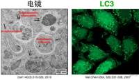
自噬共聚焦(confocal)成像拍照服务
¥2000

GenMute™ siRNA Transfection Reagent for Primary Neurons
¥1980

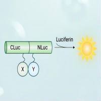
荧火素酶互补实验(Luciferase Complementation Assay, LCA)| 荧光素酶互补成像技术(Luciferase Complementation Imaging, LCI)
¥5999
相关方法

