Small Animal Imaging and Examination by Micro-CT
互联网
410
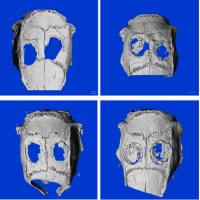
Micro-computed tomography imaging technology allows for the whole-mount investigation of skeletal structures in preclinical specimens at both fetal and postnatal time points. The imaging process is nondestructive to the specimen and can be performed at various resolutions to derive the region of interest information most relevant to individual researchers. In addition to qualitative imaging of skeletal samples, accurate and desirable metrics such as bone mineral density (BMD), discrete cortical and trabecular bone analysis, and milligrams hydroxyapatite per unit volume (mgHA/cc) are also achievable depending upon scanner platform. Additional benefits to the method include digitally archivable files, in vivo and ex vivo scanning options, and volumetric or slice-thru presentation of data in standard histological or oblique orientations. Some drawbacks to the method include long scan times at higher spatial resolutions, large file sizes, and limitation to imaging of highly dense biological structures (i.e., bone), though several groups have attempted to expand the modality to include soft tissue imaging in ex vivo specimens.