Mass Mapping of Amyloid Fibrils in the Electron Microscope Using STEM Imaging
互联网
互联网
相关产品推荐

alpha-Synuclein重组蛋白|Alpha Synuclein (A53T) Pre-Formed Fibrils (Type 1)
¥11300

Zika virus (ZIKV) (strain Zika SPH2016) ZIKV-E (Stem/anchor domain of flavivirus envelope glycoprotein E) protein (Fc Tag)
¥4520

Recombinant-Cellvibrio-japonicus-Electron-transport-complex-protein-RnfErnfEElectron transport complex protein RnfE
¥10892

Recombinant-Hordeum-vulgare-Low-molecular-mass-early-light-inducible-protein-HV60-chloroplasticLow molecular mass early light-inducible protein HV60, chloroplastic; ELIP
¥9968

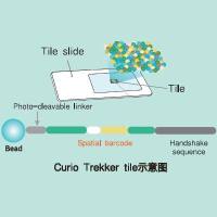
Trekker Single-Cell Spatial Mapping Kit 单细胞空间转录组分析试剂盒
询价
相关问答

