Environmental Scanning Electron Microscope Imaging of Vesicle Systems
互联网
互联网
相关产品推荐
Recombinant-Aliivibrio-salmonicida-Electron-transport-complex-protein-RnfDrnfDElectron transport complex protein RnfD
¥11900
marA/marA蛋白//蛋白/Recombinant Shigella sonnei Transcriptional activator of defense systems (marA)重组蛋白
¥69
Recombinant-Mouse-Vesicle-transport-through-interaction-with-t-SNAREs-homolog-1AVti1aVesicle transport through interaction with t-SNAREs homolog 1A Alternative name(s): Vesicle transport v-SNARE protein Vti1-like 2 Vti1-rp2
¥10710
disA/disA蛋白/Cyclic di-AMP synthase (c-di-AMP synthase) (Diadenylate cyclase)蛋白/Recombinant Mycobacterium paratuberculosis DNA integrity scanning protein DisA (disA)重组蛋白
¥69
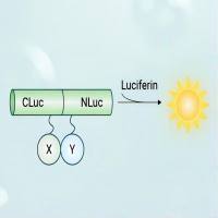
荧火素酶互补实验(Luciferase Complementation Assay, LCA)| 荧光素酶互补成像技术(Luciferase Complementation Imaging, LCI)
¥5999

