Vybrant® DyeCycle Violet Stain
互联网
1699
实验材料
Contents and storage information.
|
When stored as directed, this kit is stable for at least 6 months. |
Materials Required but Not Provided
实验步骤
For optimal DNA content cell cycle analysis, follow these guidelines:
- Eliminate cell clumps and aggregates from the cell suspension before staining
- Use 37?C for incubation with the Vybrant® DyeCycle™ Violet stain
- Hanks’ Balanced Salt Solution (HBSS) is recommended if media is not desired, however phosphate buffers are not recommended
- Do not use glass containers with this stain
- Do not wash or fix cells after staining cells with Vybrant® DyeCycle™ Violet stain
- Validate flow cytometry instrument performance on the day of use
- Use linear amplification for DNA content
- Use low flow rate for acquisition
- Collect adequate numbers of events for the intended application
- Eliminate dead cells from the DNA content analysis of living cells using a dead cell discriminating stains such as SYTOX® Green, SYTOX® Red or SYTOX® AADvanced™ dead cell stains or LIVE/DEAD® Fixable Dead cell stains such as Green, Red, Far Red, or Near-IR kits
- Compensation may be required for Alexa Fluor® 488 and PE channels
- Eliminate or correct for cell aggregates during data analysis using gating or modeling software
- Human and rodent stem cells efflux Vybrant® DyeCycle™ Violet stain, and this is the basis for the Side Population (SP) technique; the efflux can be blocked with verapamil, fumitermorgin C, or other such blocking agents, to prevent dye efflux for accurate DNA content analysis in these stem cells
Vybrant ® DyeCycle™ Violet Staining Protocol
- Remove the Vybrant® DyeCycle™ Violet stain from the refrigerator and allow the vial to equilibrate to room temperature.
- Prepare flow cytometry tubes each containing 1 mL of cell suspension in complete media at a cell concentration of 1 × 10⁶ cells/mL.
- To each tube, add 1 μL of Vybrant® DyeCycle™ Violet stain and mix well. Final stain concentration is 5 μM.
- Incubate at 37?C for 30 minutes, protected from light. Keep cells at 37?C until acquisition.
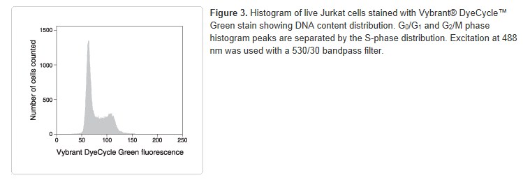
- Analyze samples without washing or fixing on a flow cytometer using ~405 nm excitation and ~440 nm emission (Figure 2). Vybrant® DyeCycle™ Violet stain may also be excited with a UV light source.