疟原虫形态 Morphology
互联网
形态:


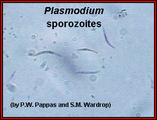
子孢子

红外期裂殖体

Pre-erythrocytic schizont in liver.
These mature in 6-14 days’ time liberation merozoites into the blood stream. Giemsa-colophonium.
×
400. Enlarged by 5.4.

环状体

P. vivax thin smear, showing early trophozoites.
The
infected red cells are enlarged and show some stippling. Giemsa.
×
1000. Enlarged by 5.4.

P. vivax thick smear, early trophozoites.
The red cells are lysed. Giemsa.
×
1000. Enlarged by 5.4.
成熟滋养体

P.vivax thin smear.
Showing late trophozoites (amoeboid stage). The infected red cells are enlarged and show marked stippling. Giemsa.
×
1000. Enlarged by 5.4.
裂殖体


P. vivax thin smear.
A mature schizont about to rupture. A clump of malarial pigment can be seen in the center. Giemsa.
×
1000. Enlarged by 5.4.

P. vivax thin smear
.
Merozoites lying free. Malarial pigment is seen as a clump on one side. Giemsa.
×
1000. Enlarged by 5.4.

间日疟原虫雄配子体

间日疟原虫雌配子体