Imaging of Vaccinia Virus Entry into HeLa Cells
互联网
互联网
相关产品推荐

Capsid L1重组蛋白|Recombinant Human Papilloma Virus type 16 (HPV 16) L1 protein (VLP)
¥3220

CHO cells 多克隆抗体 27803-1-AP
¥1350

Recombinant-Laccaria-bicolor-Protein-GET1GET1Protein GET1 Alternative name(s): Guided entry of tail-anchored proteins 1
¥10444

A33R Protein重组蛋白|Recombinant Vaccinia virus (VACV) (strain Copenhagen) A33R Protein (His Tag)
¥5820

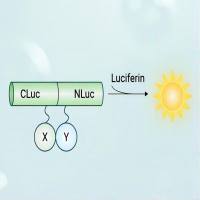
荧火素酶互补实验(Luciferase Complementation Assay, LCA)| 荧光素酶互补成像技术(Luciferase Complementation Imaging, LCI)
¥5999
相关问答

