New double-positivity tool for multi-color IHC IF
互联网
Abstract
Spatial and proteomic information are retained and varying combinations of biomarkers can be investigated, including nuclear-to-cytoplasmic expression of a single marker, colocalization of two markers in a given cellular compartment (e.g., the nucleus), and coexpression of markers in different cellular compartments, with a new double-positivity tool.

These kinds of questions can be answered in a limited way by methods that disaggregate or homogenize tissue, or that utilize conventional single-stain IHC. Simultaneous assessment of two proteins in intact FFPE tissue sections enables more accurate determination of cell state. Double-positivity assessments reveal the percentages of cells that express both, just one, or neither of two proteins. Double-positivity assessment can be used to answer many demanding analytical questions about cells in disease-related tissues, such as (i ) how many EGFR-positive cells are actively signaling, (ii ) what percentage of tumor cells are proliferating or dying, (iii ) are there any cancer stem cells present, or (iv ) how many cells have had a phospho-epitope travel across the nuclear membrane?
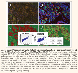
This new double-positivity tool works on multi-color immunohistochemical or immunofluorescence tissue samples by spectrally unmixing each stain or label into separate but completely co-registered component images, enabling each marker to be accurately measured even when colocalized. Furthermore, the tool allows for the isolation of immunofluorescence signals from autofluorescence, thereby dramatically improving sensitivity and signal-to-background ratio.
inForm™, an advanced yet intuitive software program with a learn-by-example interface, provides a simple, easy-to-use means of finding morphological regions of interest followed by automated cell analyses. This kind of analysis can be done on spectral images acquired from multi-label or fluorescence samples or even standard single-marker IHC images.