

影像系统

E.M. PROCESSING SCHEDULE - EPOXY RESIN Fix tissue in 2.5% glutaraldehyde in 0.1M sodium cacodylate buffer at 4oC for a minimum of 4 hours. Tissue should be cut into approx. 1mm cubes for fixing. This ...
E.M. PROCESSING SCHEDULE - EPOXY RESIN Fix tissue in 2.5% glutaraldehyde in 0.1M sodium cacodylate buffer at 4oC for a minimum of 4 hours. Tissue should be cut into approx. 1mm cubes for fixing. This ...
Fixation of Cells Cultured in Transwell DishesTranswell culture dishes are commonly used to culture cells so that the top and bottom of the cells can be exposed to different culture media conditions. ...
Chlamydomonas Fixation for Transmission Electron MicroscopySolutions: Chlamydomonas culture medium + 2% glutaraldehyde (5 ml medium + 0.9 ml 25% glutaraldehyde 100 mM sodium cacodylate pH 7.2 1% gluta ...
For routine transmission electron microscopy (TEM) it is generally accepted that specimens should be thin dry and contain molecules which diffract electrons. Biological specimens which are large and ...
EPON resin mixture for transmission electron microscopyFor Epon WPE 153: ~120 ml~60 ml~30 mlMix A:Embed 81244 ml22.1 ml11.1 mlDDSA67 ml33.3 ml16.7 mlMix B:Embed 81267 ml33.3 ml16.7 mlNMA56 ml27.8 ml13 ...
High resolution negative staining(From Valentine et al 1968. Biochemistry 7:2143-52)Rationale: For the highest resolution with negative staining there should be little or no support film but some supp ...
Preparation Of Ciliated Protozoa For Scanning Electron MicroscopyGeneral notes: The same procedures are used to fix and stain cells for SEM and for TEM. Cells can be fixed using conventional glutarald ...
Generic Fixation for Electron MicroscopyThe best way to fix a sample for electron microscopy is to follow a procedure developed and proven by others. If this is not possible this method will produce g ...
Specimen Preparation for Scanning Electron MicroscopyWe recommend consultation with one of the lab directors before preparing specimens. The methods presented here provide an overview of preparation t ...
Use of Transmission Electron Microscopy OverviewA protocol describing the use of Zeiss EM9-S transmission electron microscopy is presented.MaterialZeiss EM9-S transmission electron microscope ...
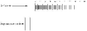
Measurement with the Light Microscope Your microscope may be equipped with a scale (called a reticule) that is built into one eyepiece. The reticule can be used to measure any planar dimension in a mi ...

Using a Counting Chamber For microbiology cell culture and many applications that require use of suspensions of cells it is necessary to determine cell concentration. One can often determine cell dens ...
Electron MicroscopesIntroductionThe introduction of the electron microscope as a tool for the biologist brought about a complete reappraisal of the micro-anatomy of biological tissues organisms and c ...
Electron MicroscopesIntroductionThe introduction of the electron microscope as a tool for the biologist brought about a complete reappraisal of the micro-anatomy of biological tissues organisms and c ...
Immunofluorescence Microscopy of tissue culture cellsThese methods are written for direct staining of filamentous actin with bodipy FL-phallicidin and indirect immunofluorescence staining of microtubu ...
Differential Interference Contrast (Nomarski DIC Hoffman Modulation Contrast) PrincipleDifferential interference microscopy requires several optical components therefore it can be very expensive to se ...
时间分辨荧光分析法(Time resolved fluoroisnmunoassayTRFIA)是近十年发展起来的一测微量分析方法,是目前最灵敏的微量分析技术其灵敏度高达10-19,较放射免疫分析(RIA)高出3个数量级。 时间分辨荧光分析法(TRFIA)实际上是在荧光分析(FIA)的基础上发展起来的,它是一种特殊的荧光分析。荧光分析的利用了荧光的波长与其激发波长的巨大差异克服了普通紫外-可见 ...
时间分辨荧光分析法(Time resolved fluoroisnmunoassayTRFIA)是近十年发展起来的一测微量分析方法,是目前最灵敏的微量分析技术其灵敏度高达10-19,较放射免疫分析(RIA)高出3个数量级。 时间分辨荧光分析法(TRFIA)实际上是在荧光分析(FIA)的基础上发展起来的,它是一种特殊的荧光分析。荧光分析的利用了荧光的波长与其激发波长的巨大差异克服了普通紫外-可见 ...
基本原理:利用半导体量子点的量子尺寸效应,即达到纳米尺度后量子点的禁带宽度随着粒度的减小而增大,禁带宽度不同在光学上表现为发射出不同颜色的光。所用利用同一光源激发后在不同大小量子点存在下会发出多种颜色的荧光。美国的量子点公司已经用这一原理,将胶体量子点与生物分析结合进入生物体,在体外用光源激发量子点发出不同颜色的光,可以进行生物分子追踪。为生命科学研究提供了一有力工具。 ...
关于丁香通
公司信息
个人用户
企业机构


