The light microscope so called because it employs visible light to detect small objects is probably the most well-known and well-used research tool in biology. Yet many students and teachers are unawa ...
USE OF THE LIGHT MICROSCOPEEach time the microscope is to be used it should be set up correctly to give a good image. Most often users forget to adjust the iris diaphragm and focus the condenser to gi ...
Use of Semi-Thin Cryosections for Light Microscopy.Semi-thin sections can be obtained from frozen blocks of cryoprotected biological material by sectioning at -90°C. The advantages for using these sec ...
Microscopes in Cell BiologyIntroductionMicroscopy has a major role in the study of cells. From the very beginning researchers have tried to develop ways of looking directly at living cells. This direc ...
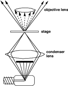
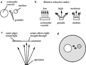
Dark Field Viewing Principle To view a specimen in dark field an opaque disc is placed underneath the condenser lens so that only light that is scattered by objects on the slide can reach the eye (fig ...
Dark Field Viewing Principle To view a specimen in dark field an opaque disc is placed underneath the condenser lens so that only light that is scattered by objects on the slide can reach the eye (fig ...
Phase Contrast MicroscopyPrincipleMost of the detail of living cells is undetectable in bright field microscopy because there is too little contrast between structures with similar transparency and no ...
Phase Contrast MicroscopyPrincipleMost of the detail of living cells is undetectable in bright field microscopy because there is too little contrast between structures with similar transparency and no ...
Microscopy with Oil ImmersionPrinciple When light passes from a material of one refractive index to material of another as from glass to air or from air to glass it bends. Light of different wavelengt ...
Microscopy with Oil ImmersionPrinciple When light passes from a material of one refractive index to material of another as from glass to air or from air to glass it bends. Light of different wavelengt ...
Tetrahymena Fixation for Transmission Electron MicroscopyPellet Tetrahymena cells in a clinical centrifuge.OPTIONAL: Suspend cells in HNMK (50 mM HEPES pH 6.9 36 mM NaCl 0.1 mM Mg acetate 1 mM KCl ) f ...
E.M. PROCESSING SCHEDULE - EPOXY RESIN Fix tissue in 2.5% glutaraldehyde in 0.1M sodium cacodylate buffer at 4oC for a minimum of 4 hours. Tissue should be cut into approx. 1mm cubes for fixing. This ...
E.M. PROCESSING SCHEDULE - EPOXY RESIN Fix tissue in 2.5% glutaraldehyde in 0.1M sodium cacodylate buffer at 4oC for a minimum of 4 hours. Tissue should be cut into approx. 1mm cubes for fixing. This ...
Fixation of Cells Cultured in Transwell DishesTranswell culture dishes are commonly used to culture cells so that the top and bottom of the cells can be exposed to different culture media conditions. ...
Chlamydomonas Fixation for Transmission Electron MicroscopySolutions: Chlamydomonas culture medium + 2% glutaraldehyde (5 ml medium + 0.9 ml 25% glutaraldehyde 100 mM sodium cacodylate pH 7.2 1% gluta ...
For routine transmission electron microscopy (TEM) it is generally accepted that specimens should be thin dry and contain molecules which diffract electrons. Biological specimens which are large and ...
EPON resin mixture for transmission electron microscopyFor Epon WPE 153: ~120 ml~60 ml~30 mlMix A:Embed 81244 ml22.1 ml11.1 mlDDSA67 ml33.3 ml16.7 mlMix B:Embed 81267 ml33.3 ml16.7 mlNMA56 ml27.8 ml13 ...
High resolution negative staining(From Valentine et al 1968. Biochemistry 7:2143-52)Rationale: For the highest resolution with negative staining there should be little or no support film but some supp ...
Preparation Of Ciliated Protozoa For Scanning Electron MicroscopyGeneral notes: The same procedures are used to fix and stain cells for SEM and for TEM. Cells can be fixed using conventional glutarald ...
Generic Fixation for Electron MicroscopyThe best way to fix a sample for electron microscopy is to follow a procedure developed and proven by others. If this is not possible this method will produce g ...


