


- ¥1700 - 4000
- GeneTex

- 美国
- GTX100685
- 2025年07月13日
- WB, ICC/IF, IHC-P, FACS, IP, PLA
- Rabbit
- Human, Mouse, Rat, Zebrafish, Bovine, Honeybee, Mosquito

企业认证
相关产品推荐更多 >
![VWF antibody [RFF-VIII R/1]](https://img1.dxycdn.com/2022/0328/360/0885999488973400453-14.jpg!wh200)
![IL2 Receptor alpha antibody [MRC OX-39] (PE)](https://img1.dxycdn.com/2022/0329/275/9974217740536020453-14.gif!wh200)
万千商家帮你免费找货
0 人在求购买到急需产品
- 详细信息
- 文献和实验
- 技术资料
- 免疫原:
Carrier-protein conjugated synthetic peptide encompassing a sequence within the center region of human SQSTM1 / P62. The exact sequence is proprietary.
- 亚型:
IgG
- 形态:
Liquid
- 保存条件:
Store as concentrated solution. Centrifuge briefly prior to opening vial. For short-term storage (1-2 weeks), store at 4ºC. For long-term storage, aliquot and store at -20ºC or below. Avoid multiple freeze-thaw cycles.
- 克隆性:
Polyclonal
- 标记物:
Unconjugated
- 适应物种:
Human, Mouse, Rat, Zebrafish, Bovine, Honeybee, Mosquito
- 保质期:
12 months from the shipping date of the product.
- 抗原来源:
Human
- 目录编号:
GTX100685
- 级别:
Primary Antibodies
- 库存:
Available
- 供应商:
GeneTex
- 宿主:
Rabbit
- 应用范围:
WB, ICC/IF, IHC-P, FACS, IP, PLA
- 浓度:
0.39 mg/ml (Please refer to the vial label for the specific concentration.)
- 靶点:
SQSTM1 / P62
- 抗体英文名:
SQSTM1 / P62 antibody [N3C1], Internal
- 抗体名:
SQSTM1 / P62 抗体 [N3C1], Internal
- 规格:
100 μl/25 μl
| 规格: | 100 μl | 产品价格: | ¥4000.0 |
|---|---|---|---|
| 规格: | 25 μl | 产品价格: | ¥1700.0 |
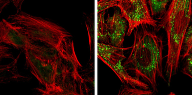
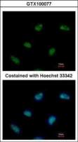
SQSTM1 antibody [N3C1], Internal detects SQSTM1 protein at autophagosome by immunofluorescent analysis.
Samples: HeLa cells mock (left) and treated with 50μM Chloroquine for 24 hr (right) were fixed in 4% PFA at RT for 15 min.
Green: SQSTM1 protein stained by SQSTM1 antibody [N3C1], Internal (GTX100685) diluted at 1:1000.
Red: Phalloidin, a F-actin marker.
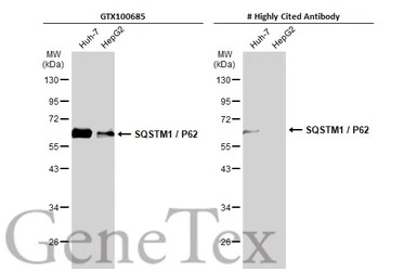
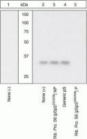
Various whole cell extracts (30 μg) were separated by 10% SDS-PAGE, and the membrane was blotted with SQSTM1 / P62 antibody [N3C1], Internal (GTX100685) diluted at 1:500. The HRP-conjugated anti-rabbit IgG antibody (GTX213110-01) was used to detect the primary antibody.
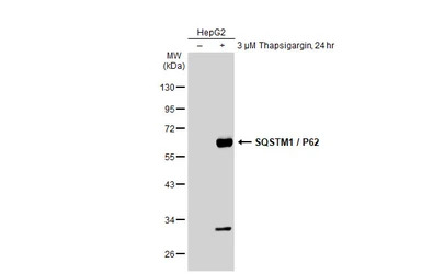
Untreated (–) and treated (+) HepG2 whole cell extracts (30 μg) were separated by 10% SDS-PAGE, and the membrane was blotted with SQSTM1 / P62 antibody [N3C1], Internal (GTX100685) diluted at 1:1000. The HRP-conjugated anti-rabbit IgG antibody (GTX213110-01) was used to detect the primary antibody.
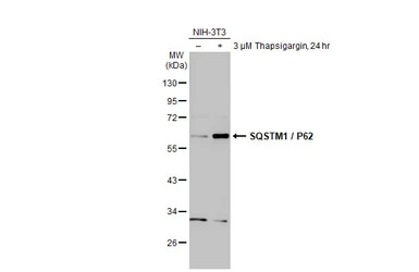
Untreated (–) and treated (+) NIH-3T3 whole cell extract (30 μg) were separated by 10% SDS-PAGE, and the membrane was blotted with SQSTM1 / P62 antibody [N3C1], Internal (GTX100685) diluted at 1:5000. The HRP-conjugated anti-rabbit IgG antibody (GTX213110-01) was used to detect the primary antibody, and the signal was developed with Trident ECL plus-Enhanced.
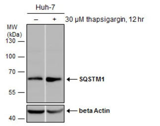
Untreated (–) and treated (+) Huh-7 whole cell extracts (30 μg) were separated by 10% SDS-PAGE, and the membrane was blotted with SQSTM1 antibody [N3C1], Internal (GTX100685) diluted at 1:1000. The HRP-conjugated anti-rabbit IgG antibody (GTX213110-01) was used to detect the primary antibody.
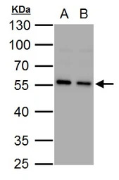
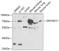
SQSTM1 antibody [N3C1], Internal detects SQSTM1 protein by western blot analysis.
A. 30 μg PC-12 whole cell lysate/extract
B. 30 μg Rat2 whole cell lysate/extract
10% SDS-PAGE
SQSTM1 antibody [N3C1], Internal (GTX100685) dilution: 1:1000
The HRP-conjugated anti-rabbit IgG antibody (GTX213110-01) was used to detect the primary antibody.
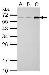
SQSTM1 antibody [N3C1], Internal detects SQSTM1 protein by western blot analysis.
A. 30 μg NIH-3T3 whole cell lysate/extract
B. 30 μg JC whole cell lysate/extract
C. 30 μg BCL-1 whole cell lysate/extract
12% SDS-PAGE
SQSTM1 antibody [N3C1], Internal (GTX100685) dilution: 1:1000
The HRP-conjugated anti-rabbit IgG antibody (GTX213110-01) was used to detect the primary antibody.
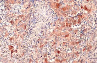
SQSTM1 / P62 antibody [N3C1], Internal detects SQSTM1 / P62 protein at cytoplasm by immunohistochemical analysis.
Sample: Paraffin-embedded human lung cancer.
SQSTM1 / P62 stained by SQSTM1 / P62 antibody [N3C1], Internal (GTX100685) diluted at 1:500.
Antigen Retrieval: Citrate buffer, pH 6.0, 15 min

Non-transfected (–) and transfected (+) HepG2 whole cell extracts (30 μg) were separated by 10% SDS-PAGE, and the membrane was blotted with SQSTM1 antibody [N3C1], Internal (GTX100685) diluted at 1:500. The HRP-conjugated anti-rabbit IgG antibody (GTX213110-01) was used to detect the primary antibody.
风险提示:丁香通仅作为第三方平台,为商家信息发布提供平台空间。用户咨询产品时请注意保护个人信息及财产安全,合理判断,谨慎选购商品,商家和用户对交易行为负责。对于医疗器械类产品,请先查证核实企业经营资质和医疗器械产品注册证情况。
 文献和实验
文献和实验Hsu CY et al., Exp Gerontol 2014 (PMID:24334178)
Tung YT et al., Mol Neurobiol 2014 (PMID:23794287)
Li JR et al., Toxicol Lett 2013 (PMID:23651616)
Hwang TI et al., Int J Oncol 2023 (PMID:37083075)
Duan C et al., Front Cell Infect Microbiol 2023 (PMID:37197204)
Cui Y et al., BMC Vet Res 2022 (PMID:35953831)
Miao CC et al., Autophagy 2022 (PMID:34470575)
Jiang B et al., Mol Med Rep 2023 (PMID:36579660)
Mostafa DK et al., Inflammopharmacology 2022 (PMID:35913649)
Kao CH et al., Autophagy 2022 (PMID:35316161)
Yaocheng Cui et al., Molecules 2022 (PMID:35630598)
Yan Guo et al., iScience 2022 (PMID:35754718)
Kuo-Hao Ho et al., Neurotherapeutics 2021 (PMID:33410111)
Tung Chao et al., Autophagy 2021 (PMID:33465003)
Martina Moras et al., Int J Mol Sci 2020 (PMID:33260618)
St?phanie Torrino et al., Elife 2021 (PMID:33884955)
Qing Yu et al., Front Pharmacol 2021 (PMID:33981237)
Valentina Naef et al., Int J Mol Sci 2021 (PMID:34445111)
Agote-Ar?n A et al., Front Cell Dev Biol 2021 (PMID:34977012)
Lee YG et al., Sci Rep 2022 (PMID:35304586)
Daussy CF et al., Autophagy 2021 (PMID:33073673)
Siadous FA et al., Autophagy 2021 (PMID:32116095)
Chung WP et al., Sci Rep 2022 (PMID:34997132)
Watanabe M et al., Molecules 2021 (PMID:35011407)
Lee CW et al., Molecules 2022;27(2)
Lin CY et al., Antioxidant 2021;10(12)
Chen WT et al., Cancers (Basel) 2021 (PMID:34439303)
Tsai CL et al., J Pers Med 2021 (PMID:34575683)
Tsai CH et al., Food Chem Toxicol 2021 (PMID:34116103)
Cheung CHY et al., J Biomed Sci 2020 (PMID:32576196)
Chen CY et al., Sci Rep 2021 (PMID:33504864)
Liu CH et al., Cancers (Basel) 2021 (PMID:33406633)
Martello A et al., EMBO Rep 2020 (PMID:32337819)
Huang HT et al., Antioxidants (Basel) 2020 (PMID:33374730)
Wong BS et al., Cancer Manag Res 2020 (PMID:32214849)
Liao CC et al., Autophagy 2018 (PMID:30081720)
Mohapatra S et al., Cell Death Differ 2020 (PMID:32483382)
Chou YJ et al., Sci Rep 2020 (PMID:32439945)
Lin SY et al., Autophagy 2018 (PMID:29171784)
Huang WT et al., Front Pharmacol 2017 (PMID:29326587)
Jensen BK et al., EMBO Mol Med 2020 (PMID:32347002)
Weng SC et al., Insect Mol Biol 2020 (PMID:32338421)
Saito Y et al., Free Radic Biol Med 2020 (PMID:32217192)
Yi H et al., Molecules 2018 (PMID:29865221)
Lin MW et al., Neurotherapeutics 2019 (PMID:31823156)
Lin MW et al., Antioxidants (Basel) 2019 (PMID:31906147)
Ho KH et al., Neurotherapeutics 2020 (PMID:31916238)
Chen JC et al., Biomolecules 2019 (PMID:31505885)
Lin TY et al., Autophagy 2019 (PMID:31612776)
Tan X et al., In Vitro Cell Dev Biol Anim 2019 (PMID:31429038)
Wang J et al., Neoplasia 2019 (PMID:31401412)
Hsieh CC et al., Br J Pharmacol 2019 (PMID:31265743)
Wang YY et al., Oral Dis 2019 (PMID:30620118)
Peng X et al., Cell Death Dis 2019 (PMID:30874544)
Lin KL et al., Free Radic Res 2018 (PMID:30693838)
Liu YH et al., Phytomedicine 2018 (PMID:30097117)
Zheng ZY et al., Sci Rep 2018 (PMID:30446677)
Ko JL et al., Environ Toxicol 2018 (PMID:30136359)
Chiu YH et al., Int J Oncol 2018 (PMID:30106130)
Yinhui Li et al., ACS OMEGA 2018;3(6)
Peng M et al., Oncogene 2018 (PMID:29930380)
Liu GT et al., FASEB J 2018 (PMID:29481305)
Chen CH et al., J Appl Physiol 2018 (Epub)
Chen CH et al., Clin Cancer Res 2017 (PMID:29222162)
Hsin MC et al., Cell Death Dis 2017 (PMID:28981104)
Huang CC et al., J Cell Sci 2014 (PMID:24860144)
Lin LC et al., J Funct Foods 2017 33()
Liu YH et al., Sci Rep 2017 (PMID:28134285)
Hai J et al., Sci Rep 2017 (PMID:28059079)
Hsin IL et al., Phytomedicine 2016 (PMID:27823620)
Lappas M et al., Reprod Sci 2017 (PMID:27638291)
Tsai JH et al., Int J Mol Sci 2016 (PMID:27527160)
Tsai WT et al., J Biol Chem 2016 (PMID:27458013)
Li H et al., Autophagy 2016 (PMID:27304991)
Hsu CY et al., Biogerontology 2016 (PMID:27230748)
Hsieh YY et al., Cell Death Discovery 2016 2()
Tseng AH et al., Free Radic Biol Med 2013 (PMID:23665396)
Chiu LY et al., PLoS One 2015 (PMID:25946033)
Liu TP et al., Anticancer Drugs 2015 (PMID:25203626)
Li KC et al., Mol Cancer 2014 (PMID:25027955)
Li JR et al., Anticancer Research 2014 (PMID:24922662)
标本。 2. 加20ul 1 N HC I ,盖紧,上下混匀。 2 - 8 ℃ 放置60± 2 分钟。 3. 加20ul 1 N NaOH, 盖紧,上下混匀。 4. 即用,或放-20/-70 ℃ 保存3 天。计算结果时乘 以稀释倍数 50 。 ( 注意:不同的标本E2F1 的水平可能有较大差异,请根据实际情况灵活掌握稀释度) 5. 细胞培养上清或组织匀浆 10 倍稀释 (410ul 的标本
Protocol for anti-HA antibody Western Blotting
of interest. 2) Transfer proteins in acrylamide gel to nylon membrane. 3) Block overnight @4℃ in PBS (or TBS, it doesn't matter) with 5% non-fat dry milk. I dilute 2.5 mg in 50 ml. 4) Wash off excess milk sol'n with PBS x3 .(important if you are re-using your antibody
Monoclonal Antibody Production Protocol
the rinse twice. Add 100 ul of blocking solution to every well, leave 1 hr at room Temp or O.N at 4°C. PRIMARY ANTIBODY Add the antibody to be tested: Sup of cells = 25 ul, mix well by pipetting up and down (10 times).serum, ascites = 1:100 and a series
 技术资料
技术资料暂无技术资料 索取技术资料

