

- 询价
- 与鼠单克隆抗体相比,兔单克隆抗体具有更高的亲和力和特异性、可识别更多表位、可识别小鼠抗原等优点,在科研和诊断等领域有着很好的表现和广泛的应用。义翘神州通过构建抗体库技术开发兔单克隆抗体,直接获得抗体基因,更有利于优秀抗体的长期保存及外源表达。
- 北京
- 2025年08月12日


万千商家帮你免费找货
0 人在求购买到急需产品
- 详细信息
- 询价记录
- 文献和实验
- 技术资料
- 提供商:
北京义翘神州科技股份有限公司
- 服务名称:
兔单克隆抗体制备服务
- 规格:
询价
与鼠单克隆抗体相比,兔单克隆抗体具有更高的亲和力和特异性、可识别更多表位、可识别小鼠抗原等优点,在科研和诊断等领域有着很好的表现和广泛的应用。义翘神州通过构建抗体库技术开发兔单克隆抗体,直接获得抗体基因,更有利于优秀抗体的长期保存及外源表达。
服务优势

兔单克隆抗体服务内容
周期: 4-6个月
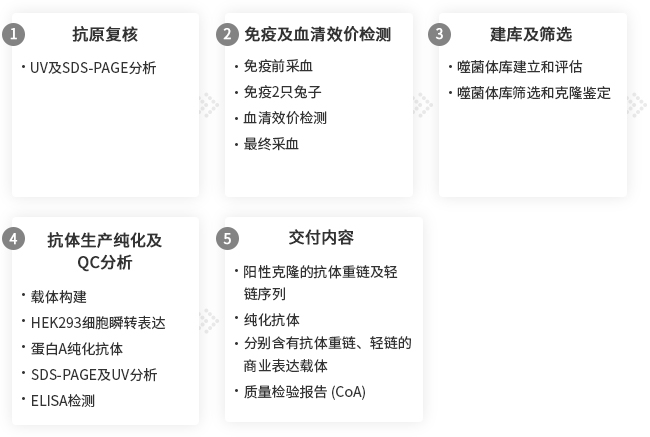
交付标准: 在ELISA检测中,纯化抗体与免疫原结合
注意: 客户可提供抗原或选择义翘神州抗原制备服务。
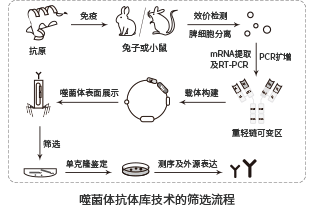
噬菌体抗体库技术开发单克隆抗体
兔单抗制备流程-噬菌体抗体库筛选基于在噬菌体展示抗体库技术方面卓越的专业知识,义翘神州已经建立了高效的兔单克隆抗体生产平台,包括制备兔抗体库和通过噬菌体展示筛选到高亲和力的抗原特异性抗体。
重组兔单克隆抗体的优势:
• 高亲和力和高特异性
• 适合检测小鼠蛋白和组织样本
• 识别更多新型表位
• 为ELISA试剂盒配对抗体开发提供多种选择
• 获得抗体基因序列,可直接用于基因工程改造
兔单克隆抗体制备服务案例
义翘神州建立了高通量兔单克隆抗体研制技术平台,通过此平台自主研发的兔单克隆抗体比传统鼠单克隆抗体具有更好的亲和力和抗原检测限。义翘神州开发的兔单抗可经过 ELISA、WB、IHC和FACS 等多种验证平台,多细胞多组织验证,提高低背景、高灵敏度抗体筛选成功率。
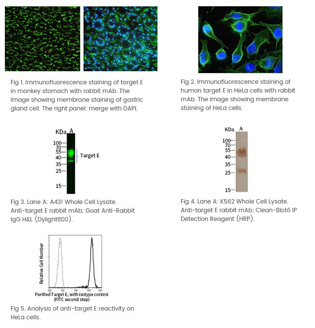
案例 1:Anti-靶点 E 兔单克隆抗体
- 多细胞、多组织验证
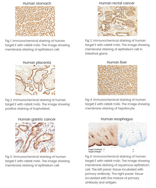
- 多应用验证
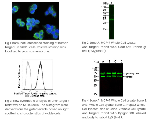
案例 2:Anti-靶点 F 兔单克隆抗体
- 多细胞、多组织验证
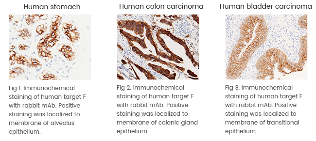
- 多应用验证
特色抗体服务
立即咨询兔单克隆抗体制备服务
扫码咨询兔单克隆抗体制备服务

(欢迎小伙伴来咨询哦! 请注明来源、姓名,单位)
下载产品相关技术资料,获取更多兔单克隆抗体制备服务相关内容!
风险提示:丁香通仅作为第三方平台,为商家信息发布提供平台空间。用户咨询产品时请注意保护个人信息及财产安全,合理判断,谨慎选购商品,商家和用户对交易行为负责。对于医疗器械类产品,请先查证核实企业经营资质和医疗器械产品注册证情况。
- 作者
- 内容
- 询问日期
 文献和实验
文献和实验- 对于不同抗原能够给出对应的免疫方案,个性化程度高,批间差异较小。 ——熊*奇 2022/5/14
- 我们的抗原之前做鼠单抗没有拿到阳性克隆,义翘的兔单抗帮忙筛选到了,很好。 ——吴*惠 2022/2/21
- 义翘帮忙筛选的兔单抗亲和力很高,而且交付序列,非常方便保存及后续表达。 ——傅* 2022/2/6
- 我的蛋白在小鼠身上免疫反应很弱,义翘的兔子免疫效果很好,而且技术人员也非常专业、nice! ——苏*蕊 2021/11/15
1975年Kohler和Milstein发现将小鼠骨髓瘤细胞与和绵羊红细胞免疫的小鼠脾细胞进行融合,形成的杂交瘤细胞既可产生抗体,又可无性繁殖,从而创立了单克隆抗体杂交瘤技术。这一技术上的突破使血清学的研究进入了一个高度精确的新纪元。 免疫细胞化学的 技术关键之一是制备特异性强、亲合力大、滴度高的特异性抗体,由于每种抗原都有几个抗原决定簇,用它免疫动物将产生对各个决定簇的抗体,即多克隆抗体。单克隆抗体则是由一个产生抗体的细胞与一个骨髓瘤细胞融合而形成的杂交廇细胞经无性繁殖而来
1. 杂交瘤阳性克隆的筛选与克隆化 在HAT培养基中生长的杂交瘤细胞,只有少数是分泌预定特异性单克隆抗体的细胞,因此,必须进行筛选和克隆化。通常采用有限稀释法进行杂交瘤细胞的克隆化培养。采用灵敏、快速、特异的免疫学方法,筛选出能产生所需单克隆抗体的阳性杂交瘤细胞,并进行克隆扩增。经过全面鉴定其所分泌单克隆抗体的免疫球蛋白类型、亚类、特异性、亲和力、识别抗原的表位及其分子量后,及时进行冻存。2. 单克隆抗体的大量制备 单克隆抗体的大量制备重要采用动物体内诱生法和体外培养法。
一、实验目的: 单克隆抗体制备是细胞免疫学的一个重要里程碑,它涵盖了细胞培养、细胞融合、免疫动物和抗体效价检测等各个方面内容。了解单克隆抗体制备的原理、主要步骤和方法。 二、实验原理: 骨髓瘤细胞在体外培养能大量无限增殖,但不能分泌特异性抗体;而抗原免疫的B淋巴细胞能产生特异性抗体,但在体外不能无限增殖。将免疫脾细胞与骨髓瘤细胞融合后形成的杂交瘤细胞,继承了两个亲代细胞的特性,既具有骨髓瘤细胞能无限制增殖的特性,又具有免疫B细胞合成和分泌特异性抗体的能力。经在HAT培养


