

- 询价
- Miltenyi Biotec

- 多产品组合
- Germany
- 2025年12月21日

企业认证
相关产品推荐更多 >


万千商家帮你免费找货
0 人在求购买到急需产品
- 详细信息
- 询价记录
- 文献和实验
- 技术资料
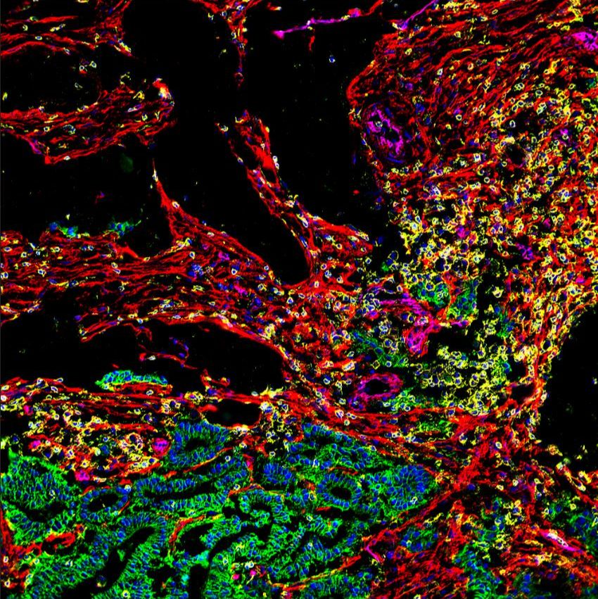
肿瘤浸润白细胞(Tumor-infiltrating Leukocytes)表型分析是了解实体肿瘤内部免疫细胞组成及其对肿瘤发展影响的重要手段。但由于TIL含量稀少,检测信号容易被淹没在背景噪声中,给TIL分析带来极大困难。
美天旎为TIL细胞表型分析特别优化的流式抗体组合令检测更容易,且完全基于重组表达的REAfinity抗体真正实现无背景的TIL流式分析:
- 基因工程令FcγR突变,无需使用FcγR阻断也可避免非特异结合;
- 只需一种IgG同型对照;
- 批次间高度一致,令结果可重复;
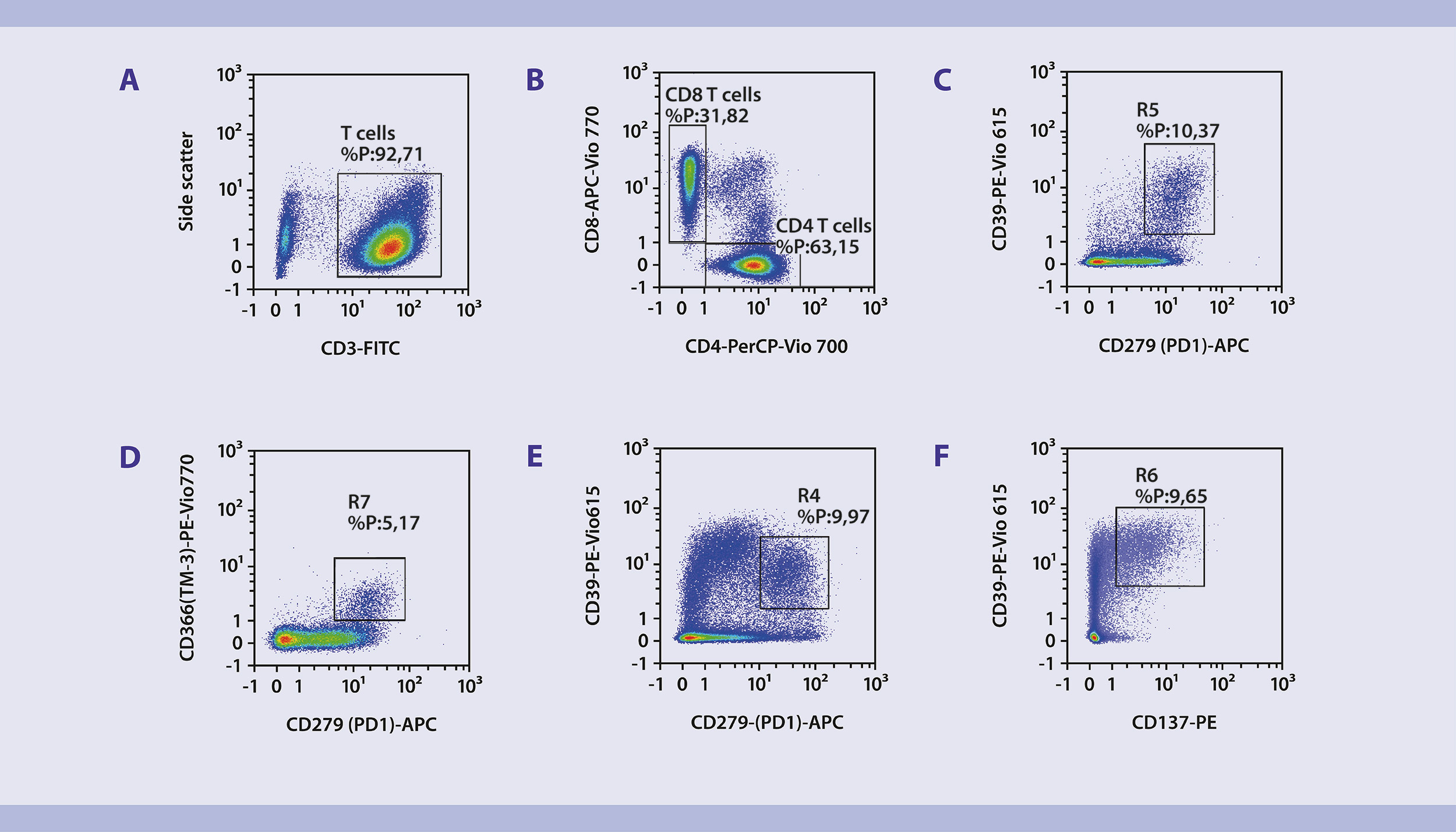
人原发卵巢肿瘤经gentleMACS Octo八通道组织解离器和Tumor Dissociation kit,human解离后,使用REAlease CD4/CD8 (TIL) MicroBeads, human 进行富集。将富集后的CD4+CD8肿瘤浸润T细胞采用如下REAfinity流式抗体组合进行免疫表型分析。
| Specificity | Clone | Purpose | Fluorochrome | Detection filter | Product No. |
| CD3 | REA613 | T cell | FITC | 525/50 | 130-113-138 |
| CD4 | REA623 | CD4+ T cell | PerCP-Vio 770 | 725/40 | 130-113-228 |
| CD8 | REA734 | CD8+ T cell | APC-Vio 770 | 785/62 | 130-110-681 |
| CD39 | REA739 | Activation marker | PE-Vio 615 | 615/20 | 130-110-656 |
| CD279(PD-1) | REA1165 | Activation marker | PE | 579/34 | 130-120-382 |
| CD366 (TIM-3) | REA635 | Activation marker | PE-Vio 770 | 785/62 | 130-121-334 |
| CD137 | REA765 | Activation marker | APC | 667/30 | 130-110-764 |
典型数据展示
Flow cytometric analysis of a human ovarian tumor sample. First, subsets of CD3+ T cells (A) were gated based on expression of CD4 and CD8 (B). The tumor-specific subpopulations CD279 (PD1)hi CD366 (TIM-)+ CD39+ CD8+ T cells and CD279 (PD1)+ CD39+ CD137+ CD4+ T cells were identified based on expression of different activation markers (PD-1, TIM-3, CD39, and CD137) within CD4+ and CD8+ T cells (C−F).
详细信息请登录美天旎官网肿瘤微环境流式检测应用页面
Immunophenotyping of tumor-specific CD4+and CD8+ T cells from a human ovarian tumor using flow cytometry
风险提示:丁香通仅作为第三方平台,为商家信息发布提供平台空间。用户咨询产品时请注意保护个人信息及财产安全,合理判断,谨慎选购商品,商家和用户对交易行为负责。对于医疗器械类产品,请先查证核实企业经营资质和医疗器械产品注册证情况。
- 作者
- 内容
- 询问日期
 文献和实验
文献和实验Production of CD4+ and CD8+ T Cell Hybridomas
T cell hybridomas are very useful tools to investigate antigen presenting cell (APC) function. They were developed based on the fusion technology that led to monoclonal antibody section. Antigen-specific primary T cells are generated
CD3/ CD4 /CD8 T cell subset counts
Description Procedure for CD3/ CD4 /CD8 T cell subset counts using CD3-FITC / CD4-PE / CD8-PECy5 antibody Procedure 1. One 5 ml round bottom tube (Falcon) is taken per blood sample to be analyzed. The tube is labeled, which includes
流式课堂 | 易被忽略的 CD4、CD8 共表达细胞——胸腺淋巴细胞
细胞的发育,成为成熟 T 细胞,然后再次随着血液循环,进入到外周淋巴器官。(图片来源于《医学免疫学》) T细胞的发育,会经历淋巴样祖细胞→祖 T 细胞→前 T 细胞→未成熟 T 细胞→成熟 T 细胞等阶段,不同阶段的 T 细胞所表达的抗原不同。根据 CD4 和 CD8 的表达来分类,胸腺中的T细胞又可以分为双阴性细胞(DN 细胞,double negative cell),双阳性细胞(DP 细胞,double positive cell)及单阳性细胞(SP 细胞,single positive
 技术资料
技术资料暂无技术资料 索取技术资料

