大家都在搜

手机验证

询价列表

暂时没有已询价产品
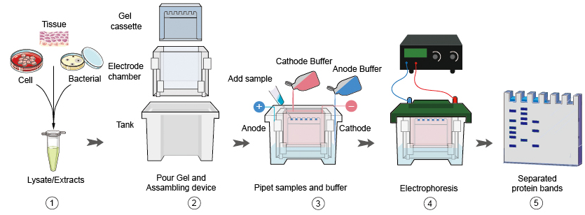
Western blot检测
BOSGENE
Western blot检测
张
2. Remove the blot from the transfer apparatus and soak in TTBS (Tween-Tris Buffered Saline: 0.1% Tween-20 in 100 mM Tris-CL [pH 7.5], 0.9% NaCl) for two rinses of 15 min. each. The blot may be marked (with pencil or India Ink) for identification at this stage if desired.
3. Block the blot with 10% nonfat dried milk (NFDM) freshly made in TTBS; rock on a rotating shaker for 15 min. at room temperature or overnight at 4°C.
4. Rinse the blot 3 times in TTBS.
5. Probe with primary antibody in TTBS/1% NFDM for 1 hr. at room temperature. Primary antibody should be diluted as specified on product data sheets. If these values are not available, use the following guidelines for initial experiments: apply antisera or ascites at 1:500 to 1:5,000, apply purified primary antibodies at a concentration of 1 ug/ml.
6. Rinse the blot three times in TTBS.
7. Probe the blot with an enzyme-linked secondary antibody (typically horseradish peroxidase or alkaline phosphatase) in TTBS/1% NFDM for 30 min. at room temperature. Review instructions included with the secondary antibody to determine the appropriate dilution to use.
8. Rinse excess secondary antibody from the blot with 3 rinses in 20-50 ml TTBS for 5 min. each. The blot is now ready for use with standard colorimetric or chemiluminescent detection reagents.
风险提示:丁香通仅作为第三方平台,为商家信息发布提供平台空间。用户咨询产品时请注意保护个人信息及财产安全,合理判断,谨慎选购商品,商家和用户对交易行为负责。对于医疗器械类产品,请先查证核实企业经营资质和医疗器械产品注册证情况。
 询价记录
询价记录
 技术资料
技术资料
博世鑫生物技术服务手册.pdf 附 (下载 0 次)