

- ¥7833
- lifeline

- 美国
- FC-0062
- 2026年03月28日


相关产品推荐更多 >
万千商家帮你免费找货
0 人在求购买到急需产品
- 详细信息
- 文献和实验
- 技术资料
- 细胞类型:
人正常原代细胞
- 物种来源:
人源
- 器官来源:
人源
- 运输方式:
干冰或液氮
- 年限:
液氮储存10年以上
- 生长状态:
冻存
- 规格:
500,000 cells/vial
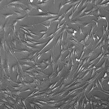
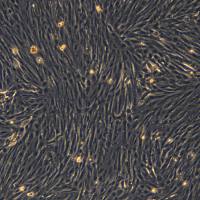
Lifeline® Human Pre-Adipocyte Cells provide an ideal culture model for the study of diabetes, obesity, metabolism, insulin sensitivity, and adipose biology.
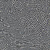
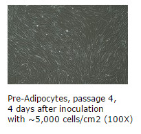
Pre-Adipocytes are derived from mature adipocytes that have been dedifferentiated, and are cryopreserved as secondary cells to ensure optimal phenotype and the highest viability and plating efficiency.
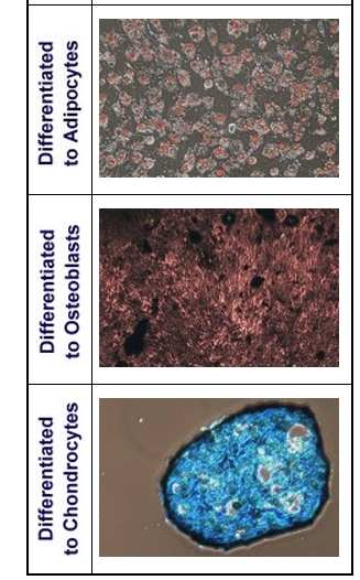
Human Pre-Adipocytes can be expanded in an undifferentiated state for future differentiation to mature Adipocytes. Lifeline® Human Pre-Adipocytes may also be differentiated down chondrogenic, and osteogenic lineages.
Lifeline® Human Pre-Adipocytes are characterized by flow cytometry to ensure the proper expression of multiple markers of mesenchymal stem cells. They are uniformly positive for CD29, CD44, CD73, CD90, CD105, and CD166. They are uniformly negative for CD14, CD31, CD34, and CD45.
Lifeline® has developed optimized media to expand the Human Pre-Adipocyte Stem Cell products in the undifferentiated state, as well as optimized media kits for inducing Adipogenesis, Chondrogenesis, and Osteogenesis. Additionally, Lifeline® provides convenient staining kits for staining the fully differentiated cells.
Pre-Adipocytes are quality tested for:
- Morphology: Normal morphology for 3 passages
- Cell Viability: Minimum 70% viability when thawed from cryopreservation
- Sterility Testing: Negative for mycoplasma. Negative for bacterial and fungal growth
- Virus Testing: Negative for HIV-1, HIV-2, HBV, and HCV by PCR
- Specific Staining: Positive* for CD29, CD44, CD73, CD90, CD105, CD166.
Negative* for CD14, CD31, CD34, CD45.
*Lifeline® defines positive expression as when greater than 95% of the cell population expresses that cell marker.
*Lifeline® defines negative expression as when less than 2% of the cell population expresses that cell marker.
风险提示:丁香通仅作为第三方平台,为商家信息发布提供平台空间。用户咨询产品时请注意保护个人信息及财产安全,合理判断,谨慎选购商品,商家和用户对交易行为负责。对于医疗器械类产品,请先查证核实企业经营资质和医疗器械产品注册证情况。
 文献和实验
文献和实验CTL 前体细胞的激活分化 CD8阳性CTI,的成熟分化由抗原驱动。与初始CD4T细胞一样,CTI。前体即初始CD8T细胞的激活和分化,也需要两个信号。第一信号来自TCR对pMHC I类复合结构的识别,提呈这一结构的,主要是非专职APC。第二信号的来源可能有两种途径:一是活化CD4 T细胞释放的细胞因子,可作用于靶细胞,使其表达协同刺激分子(如B7);二是直接由活化的CD4T细胞提供IL-2,后者与CTL前体表面的IL-2受体结合,促进其增殖分化。
Sci Adv:浙大祝赛勇团队成功建立人胰岛前体细胞高效扩增培养新体系
糖尿病是一种全球高发慢性病,严重影响着数亿人健康。目前,胰岛移植是糖尿病治疗中极具前景的一种方法,但该疗法仍面临供体来源短缺、免疫排斥等限制因素。 人多能干细胞具有自我更新和定向分化为功能性细胞的潜能。过去二十多年里,人多能干细胞分化为胰岛细胞技术取得了重大进展。然而,该分化过程经历多个中间步骤,操作难度大,极其耗时耗力。在分化过程中,胰岛前体细胞具有自我更新能力,而且利用其分化为胰岛细胞更具安全性和可控性。因此,建立胰岛前体细胞的培养体系是领域内至关重要的问题之一。但是,由于人胰岛前体细胞
文献速递|Science 子刊 :少突胶质前体细胞调控免疫反应及多发性硬化症早期脱髓鞘新机制
2025 年 4 月 2 日,由陆军军医大学牛建钦教授、万瑛教授与中山大学附属第七医院易陈菊研究员联合带领的研究团队在 Science translational medicine 上发表研究论文,首次揭示了少突胶质前体细胞(Oligodendrocyte precursor cell, OPC)在多发性硬化(Multiple Sclerosis, MS)早期病理过程中触发免疫反应级联激活,并导致快速脱髓鞘的新机制。 MS 是一种以中枢神经系统脱髓鞘为核心病理特征的自身免
 技术资料
技术资料暂无技术资料 索取技术资料

