


- ¥6400
- SouthernBiotech
- 15600-01
- 进口
- 2025年12月04日

企业认证
相关产品推荐更多 >

万千商家帮你免费找货
0 人在求购买到急需产品
- 详细信息
- 文献和实验
- 技术资料
- 供应商:
上海研卉生物
- 库存:
大量
- 规格:
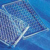
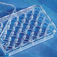
5×96孔ELISA板
产品描述:试剂盒组成(5plates):0·3mL无标记IFN-γ捕获抗体200X,0·06mL生物素标记IFN-γ检测抗体500X,0·3mLHRP标记 显色酶200X,2瓶重组人IFN-γ标准品冻干粉(2·5ng/瓶),15mL孵育液5X,300mL稀释液1X,300mL洗液20X,60mL标准 品/样品稀释液1X,60mL TMB显色液,60mL TMB反应终止液,5×96孔ELISA板,应用实验ELISA
产品特点:该试剂盒可用于培养细胞、血清、血浆中重组/天然人IFN-γ的定量检测,与人其他蛋白无交叉反应及干扰。每批次试剂盒均经过ELISA测试,以确保批间 一致性,作为一个标准的参考试剂。该试剂盒采用的是双抗体夹心法原理。无标记的人IFN-γ单克隆抗体预先在96孔板上包被固相,检测时加入标准品及待测 样本,标准品及待测样本中的IFN-γ便会与96孔板上固相抗体结合,洗板去除游离物质,依次加入生物素标记抗人IFN-γ抗体,HRP标记链酶亲合素孵 育,洗板后加入TMB显色,显色颜色深浅与样品中IFN-γ浓度正相关,在450nm处测定OD值,根据标准品和待测样品OD值可计算出IFN-γ含量
保存:所有试剂使用前稀释,稀释液中避免含有叠氮钠,以免抑制HRP酶活性,产品有效期见包装标签
Mouse Anti-Human Interferon gamma (IFN-γ) ELISA kit
Cat. No.
Form
Quantity
15600-01
Set
1
Intended Use
This set is specifically designed for quantitative determination of human interferon gamma (IFN-γ) concentrations in cell culture supernatant, serum and plasma.
Background
Human interferon gamma (IFN-γ) is a 20 or 25 kD glycoprotein that is secreted by a variety of cells, including T lymphocytes and natural killer (NK) cells1. This sole member of the type II interferon family is structurally and functionally distinct from type I members, including IFN-α and IFN-β2. IFN-γ production is primarily regulated via cytokine secretion by antigen-presenting cells (APCs)3,4. Pathogen-induced activation of APCs results in secretion of interleukin-12 and interleukin-18 that not only attract T lymphocytes and NK cells to the site of inflammation, but also induce these cells to produce IFN-γ. This up-regulation of IFN-γ promotes receptor-mediated activation of the Jak-Stat signaling pathway in target cells, leading to transcriptional modifications of target genes via specific response elements5-7.
IFN-γ mediates numerous functions in the inflammatory process. It has been shown to orchestrate specific immune cell trafficking to sites of inflammation by stimulating production of cell adhesion molecules and chemokines3,8. It promotes host response to intracellular pathogens by up-regulating cell-surface class I and II MHC molecules9,10. IFN-γ also activates microbicidal effector functions in macrophages by increasing pinocytosis and receptor-mediated phagocytosis in these cells11. Furthermore, studies have shown that IFN- may coordinate the transition from innate immunity to adaptive immunity by promoting a Th1-type response and enhancing B cell isotype switching to IgG2a12,13.
Principle of Assay
This assay incorporates a quantitative sandwich enzyme immunoassay technique. Unlabeled monoclonal antibody specific for human interferon gamma (IFN-γ) is first coated onto 96 well microplate(s). Standards and samples are then added to the wells and any IFN-γ present is captured by the immobilized antibody. After a wash to remove unbound material, biotin-labeled anti-human IFN-γ detection antibody is added to the wells, followed by a horseradish peroxidase-labeled streptavidin incubation. After another wash, TMB substrate solution is added that will result in a blue color proportional to the amount of bound IFN-γ. Color development is then quenched and intensity is measured at 450nm.
Antibody Description
Antigen Source: Human
Immunogen: Interferon gamma (IFN-γ)
Classification: Inflammation marker
Capture Antibody Detection Antibody
Host: Mouse Host: Mouse
For Research Purposes Only. Not for Diagnostic or Therapeutic Applications.
Corporate Offices: 160 Oxmoor Blvd. Birmingham, AL 35209 USA Mailing Address: P.O. Box 26221 Birmingham, AL 35260 USA
Tel: 205.945.1774 U.S. and Canada: 800.722.2255 Fax: 205.945.8768
Email: info@southernbiotech.com Website: www.southernbiotech.com
15600.doc
2 of 5
1-Mar-10
Clone: A35 Clone: B27
Isotype: IgG1 Isotype: IgG1
Research Applications
Enzyme-Linked-Immunosorbent-Assay (ELISA)
Assay Kit Components
0.3ml IFN-γ Capture Antibody: unlabeled, 200X
0.06ml IFN-γ Detection Antibody: biotin-conjugated, 1000X
0.3ml Detection Enzyme: HRP-conjugated, 200X
2 vials Standard: lyophilized recombinant human IFN-γ, 2.5ng/vial
15ml 5X Coating Buffer
300ml Assay Diluent (1X)
300ml Washing Buffer (20X)
60ml Standard/Sample Diluent
60mL TMB one component microwell substrate
60mL TMB Stop Solution
5 x 96-well ELISA plates
REAGENT PREPARATION
All reagents should be diluted immediately prior to use. Do not add sodium azide to Assay Diluent as it inhibits the activity of horseradish-peroxidase. It is recommended that all samples, controls and standards be assayed in duplicate or triplicate.
Dilute 5X Coating Buffer to 1X with distilled (DI) water. A suggested dilution (for one plate) consists of 2.2mL 5X Coating Buffer with 8.8mL of DI water.
Dilute unlabeled human IFN-γ specific Capture Antibody 200-fold with 1X Coating Buffer. A suggested dilution (for one plate) consists of 55L Capture Antibody with 10.945mL 1X Coating Buffer.
Dilute 20X Wash Buffer to 1X with DI water. A suggested dilution consists of 50mL 20X Wash Buffer with 950mL of DI water.
Reconstitute 1 vial lyophilized human IFN-γ Standard by adding 0.1ml DI water to 1 vial (25ng/mL) and gently vortex. Dilute IFN-γ Standard 100-fold by adding 5L 25ng/mL Standard to 495L Standard/Sample diluent to yield a final concentration of 250pg/mL and allow the standard to equilibrate for at least 15 minutes. Aliquot any unused standard into polypropylene vials and store at -80°C.
Dilute biotin-labeled human IFN-γ specific Detection Antibody 1000-fold with 1X Assay Diluent. A suggest dilution (for one plate) consists of 11L Detection Antibody with 10.989mL 1X Assay Diluent.
For Research Purposes Only. Not for Diagnostic or Therapeutic Applications.
Corporate Offices: 160 Oxmoor Blvd. Birmingham, AL 35209 USA Mailing Address: P.O. Box 26221 Birmingham, AL 35260 USA
Tel: 205.945.1774 U.S. and Canada: 800.722.2255 Fax: 205.945.8768
Email: info@southernbiotech.com Website: www.southernbiotech.com
15600.doc
3 of 5
1-Mar-10
Dilute horseradish peroxidase-labeled Detection Enzyme 200-fold with 1X Assay Diluent. A suggested dilution (for one plate) consists of 55L Detection Enzyme with 10.945mL 1X Assay Diluent.
Assay Procedure
1. Collect samples and prepare reagents as directed previously.
2. Add 100μL of diluted Capture Antibody solution to all wells of a provided 96-well plate. Incubate at 4oC overnight.
3. Wash entire plate 4 times with 1X Wash Buffer.
4. Add 300μL of 1X Assay Diluent per well to block non-specific binding and reduce background. Incubate at room temperature for 30 minutes.
5. Wash plate as in Step 3.
6. Prepare standard and sample dilutions and add 100μL/well of appropriate dilution to the plate. To do this, perform six two-fold serial dilutions of the 250pg/mL top standard either within the plate or in separate tubes. To dilute within the plate, add 200μl of 250pg/mL human IFN-γ Standard to row A and 100μl Standard/Sample diluent to rows B through H. Perform serial dilution by taking 100μl standard from row A and mix with row B (see below). Then take 100μl from row B and add it to row C and continue for each subsequent row. Therefore, the final human IFN-γ standard concentrations will be 250pg/mL, 125pg/mL, 62.5pg/mL, 31.3pg/mL, 15.6pg/mL, 7.8pg/mL and 3.9pg/mL. Standard/Sample Diluent alone serves as the zero standards for row H (0pg/mL). Incubate at room temperature for 2 hours.
7. Wash plate as in Step 3.
8. Add 100μL of diluted biotin-labeled Detection Antibody solution to each well and incubate at room temperature for 1 hour.
9. Wash plate as in Step 3.
10. Add 100μL of diluted HRP-labeled Detection Enzyme solution to each well and incubate at room temperature for 30 minutes.
11. Wash plate 6 times with 1X Wash Buffer. For this final wash, soak wells in Wash Buffer for 10 seconds for each wash. This will help minimize background.
12. Add 100L of TMB Substrate Solution to each well. Incubate at room temperature for 10-15 minutes. The Substrate Solution should turn blue in color.
13. Add 50L of Stop Solution to each well. The color in the wells should change from blue to yellow. If the color in the wells turns green or the color change does not appear uniform, gently tap the plate to ensure thorough mixing.
14. Determine the optical density of each well within 30 minutes using a microplate reader set to 450 nm.
Calculation of Results
Calculate the mean absorbance for each set of duplicate standards, controls and samples. Subtract the mean absorbance of the zero standards (background) from each well. Plot the standard curve on log-log graph paper, with human IFN-γ concentration on the x-axis and the mean absorbance on the y-axis. Draw the best fit straight line through the standard points. To determine the test sample human IFN-γ concentration, find the mean absorbance value of the test sample on the y-axis and draw a horizontal line
For Research Purposes Only. Not for Diagnostic or Therapeutic Applications.
Corporate Offices: 160 Oxmoor Blvd. Birmingham, AL 35209 USA Mailing Address: P.O. Box 26221 Birmingham, AL 35260 USA
Tel: 205.945.1774 U.S. and Canada: 800.722.2255 Fax: 205.945.8768
Email: info@southernbiotech.com Website: www.southernbiotech.com
15600.doc
4 of 5
1-Mar-10
to the standard curve. At the point of intersection, draw a vertical line to the x-axis and read the IFN-γ concentration. If samples were diluted, multiply by the appropriate dilution factor. Computer based curve-fitting software may be preferred. If a test sample’s O.D. value falls outside the linear portion of the standard curve, the test sample should be reanalyzed at an alternate dilution as appropriate.
Trouble Shooting
General technical hints:
To avoid cross-contamination, change pipette tips between additions of each standard level, between sample additions and between reagent additions. Also, use separate reservoirs for each reagent.
When using an automated plate washer, adding a 30 second soak period following the addition of wash buffer and/or rotating the plate 180 degrees between wash steps may improve assay precision.
During incubation steps, shaking the plates may increase sensitivity.
Substrate Solution should remain colorless until added to the plate. Keep Substrate Solution protected from light. Substrate Solution should change from colorless to gradations of blue upon addition.
Stop Solution should be added to the plate in the same order as the Substrate Solution.
The color development in the wells will turn from blue to yellow upon addition of the Stop Solution. Wells that turn green in color indicate that the Stop Solution has not mixed thoroughly with the Substrate Solution.
Poor Signal
Avoid sodium azide in wash buffers as it inhibits the enzymatic activity of HRP
Verify that appropriate antibody pairs were used
Inadequate reagent volumes added to wells
Incorrect incubation times and/or temperature
Poor Standard Curve or Precision
Improper standard handling and/or dilution
Inadequate mixing of reagents
Inadequate aspiration and/or washing of wells
References
1. Gray, P.W. et al. (1982) Nature. 295(5849): 503-8.
2. Fleckner, J., Rasmussen, H.H. and Justesen, J. (1991) Proc. Natl. Acad. Sci. 88(24): 11520-4.
3. Boehm, U. et al. (1997) Annu. Rev. Immunol. 15:749-795.
4. Zamanian-Daryoush, M. et al. (2000) Mol. Cell. Biol. 20:1278-1290.
5. Subramaniam, P. S. et al. (2001) Cytokine. 15:175-187.
6. Meraz, M. A. et al. (1996) Cell. 84:431-442.
7. Kotenko S.V. and Pestka, S. (2000) Oncogene. 19(21): 2557-65.
8. Schroder, K. et al. (2004) J. Leuk. Biol. 75: 163-89.
For Research Purposes Only. Not for Diagnostic or Therapeutic Applications.
Corporate Offices: 160 Oxmoor Blvd. Birmingham, AL 35209 USA Mailing Address: P.O. Box 26221 Birmingham, AL 35260 USA
Tel: 205.945.1774 U.S. and Canada: 800.722.2255 Fax: 205.945.8768
Email: info@southernbiotech.com Website: www.southernbiotech.com
15600.doc
5 of 5
1-Mar-10
9. Mach, B. et al. (1996) Annu. Rev. Immunol. 14: 301-331.
10. Anderson, S. L. et al. (1994) J. Exp. Med. 180: 1565-1569.
11. Decker, T. et al. (2002) J. Clin. Invest. 109: 1271-1277.
12. Huang, S. et al. (1993) Science. 259: 1742-1745.
13. Collins, J. T. and Dunnick, W. A. (1993) Int. Immunol. 5: 885-891
风险提示:丁香通仅作为第三方平台,为商家信息发布提供平台空间。用户咨询产品时请注意保护个人信息及财产安全,合理判断,谨慎选购商品,商家和用户对交易行为负责。对于医疗器械类产品,请先查证核实企业经营资质和医疗器械产品注册证情况。
 文献和实验
文献和实验Warnings And Limitations This product is for research use only. Not intended for any animal or human therapeutic or diagnostic use unless otherwise stated. Follow appropriate laboratory guidelines. This product contains 0.02% sodium azide
RAT/MOUSE GROWTH HORMONE ELISA KIT
实验原理 This assay is a Sandwich ELISA based, sequentially, on: 1) capture of rat or mouse Growth Hormone molecules from samples to the wells of a microtiter plate coated by a pre-titered amount of anti-Growth Hormone
Isolation of c-Kit+ Human Amniotic Fluid Stem Cells from Second Trimester
to obtain c-Kit+ human AFS cells, starting from second trimester amniocentesis samples.
 技术资料
技术资料暂无技术资料 索取技术资料

