


- 询价
- 2026年05月29日

企业认证


万千商家帮你免费找货
0 人在求购买到急需产品
- 详细信息
- 文献和实验
- 技术资料
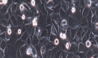
- 细胞形态:
上皮样
- 年限:
15 years
- 运输方式:
冻存运输
- 生长状态:
贴壁生长
- 器官来源:
骨
- 物种来源:
人
- 是否是肿瘤细胞:
1
- 库存:
大量
- 相关疾病:
骨肉瘤
- ATCC Number:
HTB-96™
| Designations: | U-2 OS | ||
| Depositors: | Hellstrom | ||
| Biosafety Level: | 1 | ||
| Shipped: | frozen | ||
| Medium & Serum: | See Propagation | ||
| Growth Properties: | adherent | ||
| Organism: | Homo sapiens | ||
| Morphology: | epithelial |
||
| Source: | Organ: bone Disease: osteosarcoma |
||
| Cellular Products: | osteosacoma derived growth factor (ODGF) | ||
| Permits/Forms: | In addition to the MTA mentioned above, other ATCC and/or regulatory permits may be required for the transfer of this ATCC material. Anyone purchasing ATCC material is ultimately responsible for obtaining the permits. Please click here for information regarding the specific requirements for shipment to your location. | ||
| Applications: | transfection host | ||
| Receptors: | insulin-like growth factor I (IGF-I); insulin-like growth factor II (IGF II) | ||
| Antigen Expression: | Blood Type A; Rh+; HLA A2, Aw30, B12, Bw35, B40(+/-) | ||
| DNA Profile (STR): | Amelogenin: X CSF1PO: 13 D13S317: 13 D16S539: 11,12 D5S818: 11 D7S820: 11,12 THO1: 6,9.3 TPOX: 11,12 vWA: 14,18 |
||
| Cytogenetic Analysis: | Cell line U-2 OS is chromosomally highly altered, with chromosome counts in the hypertriploid range. We did not find the hypodiploid cell population described by J. Ponten, et al., Instead, most of the population has slightly higher counts than first described. Very few normal chromosomes are present, but a high number of stable marker chromosomes are identified., Different chromosomal rearrangements involving the same chromosomes (N1, N7, N9, and N11 particularly), are seen. Twenty-two markers are found including: t(9qter--->9q21::1p36--->1p::?), 7p+, iso(17q), t(15q;?), 4q+, del(3)(q21), 5q(aberrant) and others. [22509 ] | ||
| Isoenzymes: | AK-1, 1 ES-D, 1 G6PD, B GLO-I, 2 PGM1, 2 PGM3, 1 |
||
| Age: | 15 years | ||
| Gender: | female | ||
| Ethnicity: | Caucasian | ||
| Comments: | J. Ponten and E. Saksela derived this line (originally 2T) in 1964 from a moderately differentiated sarcoma of the tibia of a 15 year old girl. Viruses were not detected during co-cultivation with WI-38 cells or in CF tests against SV40, RSV or adenoviruses. Mycoplasma contamination was detected and eliminated in 1972. |
||
| Propagation: | ATCC complete growth medium: The base medium for this cell line is ATCC-formulated McCoy's 5a Medium Modified, Catalog No. 30-2007. To make the complete growth medium, add the following components to the base medium: fetal bovine serum to a final concentration of 10%. Temperature: 37.0°C |
||
| Subculturing: | Subcultivation Ratio: A subcultivation ratio of 1:3 to 1:6 is recommended Medium Renewal: 2 to 3 times per week Remove medium, and rinse with 0.25% trypsin, 0.03% EDTA solution. Remove the solution and add an additional 1 to 2 ml of trypsin-EDTA solution. Allow the flask to sit at room temperature (or at 37C) until the cells detach. Add fresh culture medium, aspirate and dispense into new culture flasks. |
||
| Preservation: | Culture medium, 95%; DMSO, 5% | ||
| Related Products: | recommended serum:ATCC 30-2020 | ||
| References: | 22237: Heldin CH, et al. A human osteosarcoma cell line secretes a growth factor structurally related to a homodimer of PDGF A-chains. Nature 319: 511-514, 1986. PubMed: 3456080 22509: Ponten J, Saksela E. Two established in vitro cell lines from human mesenchymal tumours. Int. J. Cancer 2: 434-447, 1967. PubMed: 6081590 23011: Raile K, et al. Human osteosarcoma (U-2 OS) cells express both insulin-like growth factor-I (IGF-I) receptors and insulin-like growth factor-II/mannose-6- phosphate (IGF-II/M6P) receptors and synthesize IGF-II: autocrine growth stimulation by IGF-II via the IGF-I receptor. J. Cell. Physiol. 159: 531-541, 1994. PubMed: 8188767 32288: Landers JE, et al. Translational enhancement of mdm2 oncogene expression in human tumor cells containing a stabilized wild-type p53 protein. Cancer Res. 57: 3562-3568, 1997. PubMed: 9270029 32308: Moradpour D, et al. Characterization of cell lines allowing titghtly regulated expression of heapatitis C virus core protein. Virology 222: 51-63, 1996. PubMed: 8806487 |
||
风险提示:丁香通仅作为第三方平台,为商家信息发布提供平台空间。用户咨询产品时请注意保护个人信息及财产安全,合理判断,谨慎选购商品,商家和用户对交易行为负责。对于医疗器械类产品,请先查证核实企业经营资质和医疗器械产品注册证情况。
 文献和实验
文献和实验这类研究一般用于预后研究,大概是怎么做的呢?还是用个实例来说最最清晰。 确定研究人群:首先我们都知道 AFP(甲胎蛋白)可用于肝癌的诊断,于是我们假设 AFP 与肺癌的预后有关。那么我们的研究人群就可以按照基线 AFP 的水平将病人分成若干组,这就是若干个 Cohort(队列)。 数据收集:可以回顾性的收集手头可以接触到的一切病例资料,越多越好。 随访:病例资料收集完毕后,我们可以对这若干组人群进行随访,随访设立一个终点,例如死亡。 结局分析:随访一段时间后,可以得到这些组别的 OS(总生存
激光诱导 DNA 损伤后 U2OS 细胞的活细胞成像 双链 DNA 断裂是对 DNA 损伤最有害的形式之一。损伤之后,细胞中的 DNA 损伤反应(DDR)通路被触发,诱导 DDR 因子募集到断裂位点,并启动细胞周期检查点信号传导和 DNA 修复活性的调控。精准的信号转导和断裂位点修复对于细胞的生存和防止致癌突变至关重要。因此,了解 DNA 修复过程中的相关机制尤为关键。在此应用中,我们使用 FV3000 共聚焦显微镜,研究了 U2OS 细胞(人骨肉瘤上皮细胞)DNA 修复蛋白在激光诱导损伤
文献速递:个性化循环肿瘤 DNA 可成为免疫治疗疗效新型biomarker
性乳腺癌 (TNBC)、高级别浆液性卵巢癌 (HGSOC)、恶性黑色素瘤 (MM) 和混合实体瘤 (MST) 在内的肿瘤前瞻性2期临床研究 (NCT02644369),以评估ctDNA检测在预测抗PD-1抗体治疗效果方面的能力。通过对94名患者的316份血浆样本进行ctDNA水平测定,发现基线 ctDNA 浓度与无进展生存期、总生存期、临床反应和临床获益相关。具体表现为基线ctDNA低于中位基线ctDNA的患者总生存率(OS)和无进展生存率(PFS)更长(HR=0.49;HR=0.54)。且基线ctDNA水平低
 技术资料
技术资料暂无技术资料 索取技术资料

