

- 询价
- abbelight
- SAFe 360
- 法国
- 2026年06月08日

企业认证
相关产品推荐更多 >

万千商家帮你免费找货
0 人在求购买到急需产品
- 详细信息
- 询价记录
- 文献和实验
- 技术资料
- 保修期:
面议
- 供应商:
Quantum量子科学仪器贸易(北京)有限公司
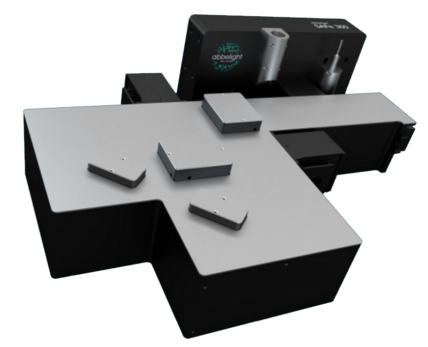
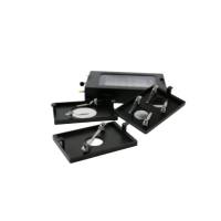
3D单分子荧光成像系统-SAFe 360
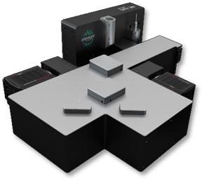
3D单分子荧光成像系统-SAFe 360
+ 成像模式:PALM、STORM、PAINT、smFRET 、SPT
+ 光源模式:Epi、TIRF、HILO
+ 超高分辨率:15 nm的XYZ轴分辨率
+ 超大视野:200 × 200 μm2的视野
+ 一次可同时采集1.2 μm深度图像信息
+ 大图像深度:10 μm
+ 实时漂移矫正
+ 多四色同时成像
+ 活细胞成像模式
| 加装 TIRF PALM STORM SPT smFRET ...... |
兼容 Confocal Spinning-Desk Widefield SIM STED |
 |
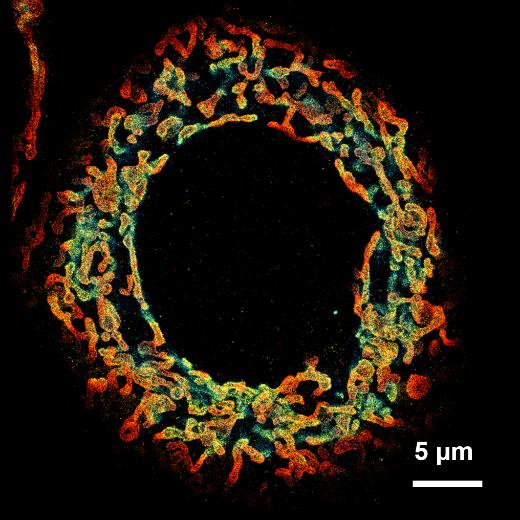 3D线粒体结构 |
 核孔复合物 |
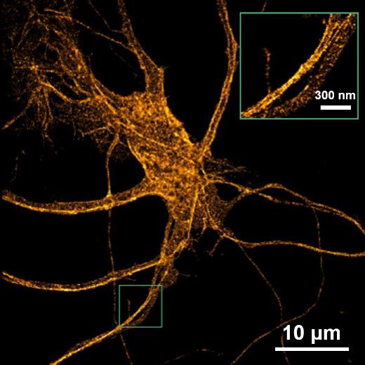 老鼠海马神经元 |
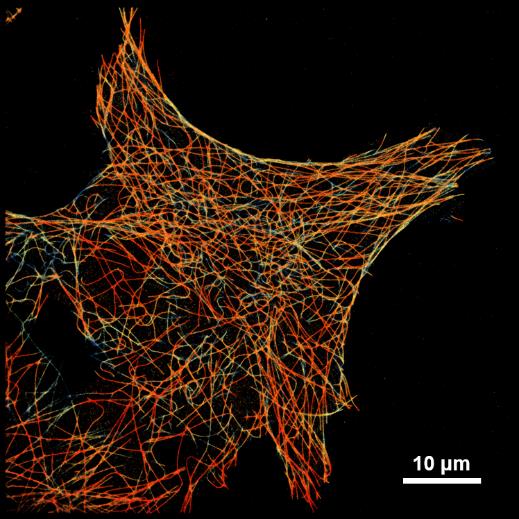 微管蛋白网络 |
配套试剂
| Smart kit • 10 doses per box • 200 µL per dose • 30 sec prepartion • 2 months in a fridge • 2 weeks on sample |
Compatible dyes • Atto 488, WGA-AF®488 • AF®532, CF®532, Cy3b • AF®555, AF®594, CF®555, AF®568, CF®568, Cy5, MemBriteTM 568, TMR • AF®647, CF®647, AF®680, CF®680, MemBriteTM 640, Actin-stain 670, SiR647 |
[1] Radhakrishnan, A. V., et al. "Single-Protein Tracking to Study Protein Interactions During Integrin-Based Migration." The Integrin Interactome. Humana, New York, NY, (2021). 85-113.
[2] Jouchet, Pierre, et al. "Nanometric axial localization of single fluorescent molecules with modulated excitation." Nature Photonics (2021): 1-8.
[3] Pernier, Julien, et al. "Myosin 1b flattens and prunes branched actin filaments." Journal of cell science 133.18 (2020).
[4] Jimenez, Angélique, Karoline Friedl, and Christophe Leterrier. "About samples, giving examples: optimized single molecule localization microscopy." Methods 174 (2020): 100-114.
[5] Mau, Adrien, et al. "Fast scanned widefield scheme provides tunable and uniform illumination for optimized SMLM on large fields of view." bioRxiv (2020).
[6] Orre, Thomas, et al. "Molecular motion and tridimensional nanoscale localization of kindlin control integrin activation in focal adhesions." bioRxiv (2020).
[7] Cabriel, Clément, et al. "Combining 3D single molecule localization strategies for reproducible bioimaging." Nature communications 10.1 (2019): 1980.
[8] Woodhams, Stephen G., et al. "Cell type–specific super-resolution imaging reveals an increase in calcium-permeable AMPA receptors at spinal peptidergic terminals as an anatomical correlate of inflammatory pain." Pain 160.11 (2019): 2641-2650.
[9] Belkahla, Hanen, et al. "Carbon dots, a powerful non-toxic support for bioimaging by fluorescence nanoscopy and eradication of bacteria by photothermia." Nanoscale Advances (2019).
[10] Denis, Kevin, et al. "Targeting Type IV pili as an antivirulence strategy against invasive meningococcal disease." Nature microbiology 4.6 (2019): 972.
[11] Szabo, Quentin, et al. "TADs are 3D structural units of higher-order chromosome organization in Drosophila." Science advances 4.2 (2018): eaar8082.
[12] Boudjemaa, Rym, et al. "Impact of bacterial membrane fatty acid composition on the failure of daptomycin to kill Staphylococcus aureus." Antimicrobial agents and chemotherapy 62.7 (2018): e00023-18.
[13] Culley, Siân, et al. "Quantitative mapping and minimization of super-resolution optical imaging artifacts." Nature methods 15.4 (2018): 263.
[14] Berger, Stephen L., et al. "Localized myosin II activity regulates assembly and plasticity of the axon initial segment." Neuron 97.3 (2018): 555-570.
[15] Cabriel, Clément, et al. "Aberration-accounting calibration for 3D single-molecule localization microscopy." Optics letters 43.2 (2018): 174-177.
[16] Bouissou, Anaïs, et al. "Podosome force generation machinery: a local balance between protrusion at the core and traction at the ring." ACS nano 11.4 (2017): 4028-4040.
[17] Sellés, Julien, et al. "Nuclear pore complex plasticity during developmental process as revealed by super-resolution microscopy." Scientific reports 7.1 (2017): 14732.
[18] Bourg, Nicolas, et al. "Direct optical nanoscopy with axially localized detection." Nature Photonics 9.9 (2015): 587.
用户单位

风险提示:丁香通仅作为第三方平台,为商家信息发布提供平台空间。用户咨询产品时请注意保护个人信息及财产安全,合理判断,谨慎选购商品,商家和用户对交易行为负责。对于医疗器械类产品,请先查证核实企业经营资质和医疗器械产品注册证情况。
- 作者
- 内容
- 询问日期
 文献和实验
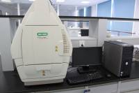
文献和实验IN Cell Analyzer 2000:斑马鱼自动化全孔成像
相关专题 细胞培养相关用品 小小斑马鱼对人类科研的大大贡献 摘要 : IN Cell Analyzer 2000是一种全新的高性能成像系统,灵活地设计足以应对多种需求——从全自动显微镜到自动化的高内涵筛选。在此展示了IN Cell Analyzer 2000在斑马鱼(药物开发和毒性研究的模式生物 )分析应用中的全控成像能力。 作者 :1 Ahmad Yekta*, 1 Zahra Masoumi, 2 Albie
子成像经历了惊人的演变,现在通过使用单分子作为超高频发射器达到定点。“这个发展太棒了,在研究单分子几乎20年后,”他说。 2008及展望 自从2006年STORM和PALM问世以来,研究者兴奋地研究改进和新应用软件。2008年也不例外。Lippincott-Schwartz的小组把PALM和单粒子失踪结合起来探测肝细胞膜蛋白的运动。《科学》中,Zhuang的小组展示了3D STORM成像,比三维空间衍射极限空间分辨率好十倍,并使用此方法在猴肾细胞中成像微管和其他分子结构,后来扩展此方法为整个细胞
器,油压式微量注射器。 3.选配件:电动注射器、电动可编程精确注射器。 4.选配件:CCTV闭路电视监视系统。 5.选配件:MHW-3水压式电生理显微操作器。 用于单分子荧光的TIRF系统 1.TIRF显微术具有可提供发微弱荧光分子的极高信噪 比图像的优点,使用尼康独创的可彻底消除入射杂散光的“噪声消除器"机构之后,该优点得到极大的增强。 2.专用的60X/100X平场复消色差物镜(油镜)数值孔径达到1.49,温度较正环最大
 技术资料
技术资料暂无技术资料 索取技术资料

