


Miltenyi货号130-090-485抗生物素磁珠Ant
i-Biotin MicroBeads上海睿安生物Miltenyi美天旎Miltenyi官方正式授权代理商19121878899- ¥7586.82
- Miltenyi Biotec
- 130-090-485
- Made in Germany
- 2026年11月10日

企业认证
相关产品推荐更多 >

美天旎Miltenyi货号200-073-104现货CliniMACS® Tubing Set,CE上海睿安生物13611631389
¥20473.34
美天旎Miltenyi货号130-041-407预分离过滤器Pre-Separation Filters(30µm)上海睿安生物13611631389
¥2352.66
美天旎Miltenyi货号130-093-545全血柱试剂盒(Whole Blood Column Kit)上海睿安生物19121878899美天旎Miltenyi官方正式授权
¥1978.63
Miltenyi Biotec货号130-139-368热销Human CD4 MicroBeads UltraPure上海睿安生物Miltenyi美天旎Miltenyi官方正式授权代理商19121878899
¥8993.67
Miltenyi货号130-104-075小鼠脾脏和淋巴结单细胞悬液中分离未经标记的CD8a⁺ T细胞(mouse CD8a⁺ T Cell Isolation Kit)上海睿安生物Miltenyi美天旎Miltenyi官方正式授权代理商19121878899
¥9286.35
万千商家帮你免费找货
0 人在求购买到急需产品
相关阅读
Dove 医学期刊 APC 折后约 1.95w,中国大陆作者专享AJE 润色 84 折,送京东卡等福利,优惠码 DXYAJE五大应用案例:Mustang Q 膜层析应用全解析1 个小工具,一次性搞定流程图、质粒图谱和信号通路图- 详细信息
- 文献和实验
- 技术资料
- 英文名:
Anti-Biotin MicroBeads
- 保质期:
1年
- 保存条件:
2-8°C
- 库存:
30⁺盒
- 供应商:
上海睿安生物19121878899
- 规格:
1╳10⁹ total cells/盒
Miltenyi Biotec货号130-090-485抗生物素磁珠Anti-Biotin MicroBeads上海睿安生物Miltenyi美天旎Miltenyi官方正式授权代理商19121878899
Anti-Biotin MicroBeads
Anti-Biotin MicroBeads have been developed for indirect magnetic labeling and separation of cells or other biological materials that are labeled with a biotinylated primary antibody or ligand, or with a cocktail of biotinylated antibodies. When working with debris-rich starting material and low frequency samples we recommend Anti-Biotin MicroBeads UltraPure to benefit from high specific enrichment with low background of debris and dead cells.
Specifications for Anti-Biotin MicroBeads
Overview
Anti-Biotin MicroBeads have been developed for indirect magnetic labeling and separation of cells or other biological materials that are labeled with a biotinylated primary antibody or ligand, or with a cocktail of biotinylated antibodies. When working with debris-rich starting material and low frequency samples we recommend Anti-Biotin MicroBeads UltraPure to benefit from high specific enrichment with low background of debris and dead cells.
Detailed product information
Background information
Anti-Biotin MicroBeads UltraPure have been especially developed for highly efficient separation of cells from debris-rich starting material. The biotinylated molecule is recognized by a monoclonal anti-biotin antibody coupled to MicroBeads. Anti-Biotin MicroBeads and Anti-Biotin MicroBeads UltraPure have the advantage of not binding to free biotin, which is often present in culture media. This feature provides the most sensitive labeling and separation of cells. With UltraPure MicroBeads we offer an ideal solution for ultrapure cell separation of cells even from debris-rich samples or other low frequency biological starting materials.
Applications
Anti-Biotin MicroBeads are ideal for positive selection, even of cells with low antigen expression, or for depletion. Anti-Biotin MicroBeads have been used for the separation of murine αE-CD25+ and αE-CD25- T cell subsets1 and for the isolation of CD45.2+ cells from the bone marrow of chimeric mice2. They have also been used for the enrichment of human CD94+ NK cells3. Anti-Biotin MicroBeads UltraPure have been especially designed for high-efficient cell isolation even from debris-rich samples and starting materials with increased amount of dead cells.
Columns
For positive selection: MS, LS, XS, or autoMACS® Columns. For depletion: LD, D, or autoMACS Columns.
Product data and images for Anti-Biotin MicroBeads
Figure 1
Separation of CD3+ cells from human PBMCs using a biotinylated CD3 antibody, Anti-Biotin MicroBeads, and an MS Column.
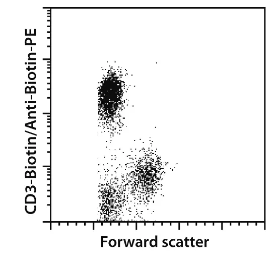
Image: PBMCs before separation
Open image in new tab
Separation of CD3+ cells from human PBMCs using a biotinylated CD3 antibody, Anti-Biotin MicroBeads, and an MS Column.
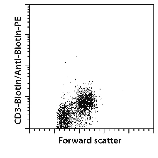
Image: CD3- cells
Open image in new tab
Separation of CD3+ cells from human PBMCs using a biotinylated CD3 antibody, Anti-Biotin MicroBeads, and an MS Column.
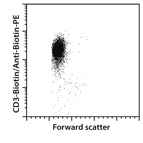
Image: CD3+ cells
Open image in new tab
Separation of CD3+ cells from human PBMCs using a biotinylated CD3 antibody, Anti-Biotin MicroBeads, and an MS Column.
Figure 2
Human CLEC9a+ cells were isolated from a debris-rich sample of peripheral blood mononuclear cells (PBMCs) using Anti-CLEC9a- Biotin, Anti-Biotin MicroBeads UltraPure, two MS Columns, and a MiniMACS™ Separator. Cells were stained with Anti-Biotin-PE (# 130-090-756), CD141 (BDCA-3)-APC (# 130-090-907), and Propidium Iodide Solution (# 130-093-233). Cells were analyzed after gating on viable lymphoid cells.
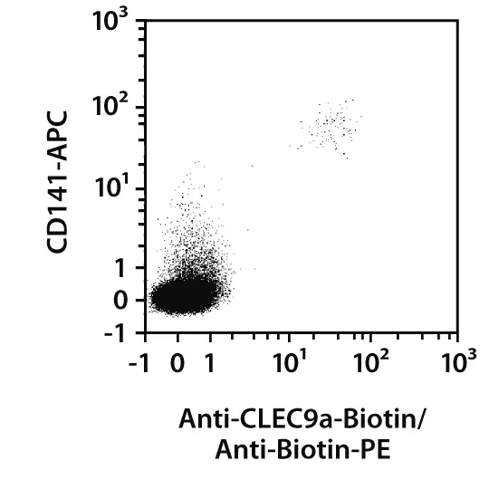
Image: Before separation
Open image in new tab
Human CLEC9a+ cells were isolated from a debris-rich sample of peripheral blood mononuclear cells (PBMCs) using Anti-CLEC9a- Biotin, Anti-Biotin MicroBeads UltraPure, two MS Columns, and a MiniMACS™ Separator. Cells were stained with Anti-Biotin-PE (# 130-090-756), CD141 (BDCA-3)-APC (# 130-090-907), and Propidium Iodide Solution (# 130-093-233). Cells were analyzed after gating on viable lymphoid cells.
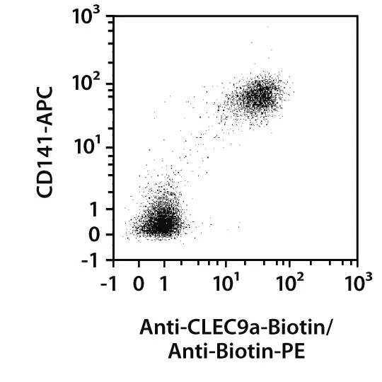
Image: After separation
Open image in new tab
Human CLEC9a+ cells were isolated from a debris-rich sample of peripheral blood mononuclear cells (PBMCs) using Anti-CLEC9a- Biotin, Anti-Biotin MicroBeads UltraPure, two MS Columns, and a MiniMACS™ Separator. Cells were stained with Anti-Biotin-PE (# 130-090-756), CD141 (BDCA-3)-APC (# 130-090-907), and Propidium Iodide Solution (# 130-093-233). Cells were analyzed after gating on viable lymphoid cells.
Citations for Anti-Biotin MicroBeads
Publications
Gastpar, R. et al. (2004) The cell surface-localized heat shock protein 70 epitope TKD induces migration and cytolytic activity selectively in human NK cells. J Immunol 172: 972-980
Lang, R. et al. (2003) SOCS3 regulates the plasticity of gp130 signaling. Nat Immunol 4: 546-550
Lehmann, J. et al. (2002) Expression of the integrin alpha Ebeta 7 identifies unique subsets of CD25+ as well as CD25- regulatory T cells. Proc. Natl. Acad. Sci. U.S.A. 99: 13031-13036

已经开发的抗生物素磁珠通过间接磁性标记分选经生物素一抗和配体标记的细胞或者其它生物材料。当起始样本中细胞碎片较多或者细胞含量很少,推荐使用超纯抗生物素磁珠。该试剂盒获得高特异性,低背景的细胞。
相关产品:
▪ MS分选柱(Miltenyi货号130-042-201)
▪ LS分选柱(Miltenyi货号130-042-401)
▪ 磁力架(Miltenyi货号130-042-303)
▪ MS分选磁极(Miltenyi货号130-042-102)
风险提示:丁香通仅作为第三方平台,为商家信息发布提供平台空间。用户咨询产品时请注意保护个人信息及财产安全,合理判断,谨慎选购商品,商家和用户对交易行为负责。对于医疗器械类产品,请先查证核实企业经营资质和医疗器械产品注册证情况。
 文献和实验
Miltenyi Biotec货号130-090-485抗生物素磁珠Anti-Biotin MicroBeads上海睿安生物Miltenyi美天旎Miltenyi官方正式授权代理商19121878899
Anti-Biotin MicroBeads
Anti-Biotin MicroBeads have been developed for indirect magnetic labeling and separation of cells or other biological materials that are labeled with a biotinylated primary antibody or ligand, or with a cocktail of biotinylated antibodies. When working with debris-rich starting material and low frequency samples we recommend Anti-Biotin MicroBeads UltraPure to benefit from high specific enrichment with low background of debris and dead cells.
Specifications for Anti-Biotin MicroBeads
Overview
Anti-Biotin MicroBeads have been developed for indirect magnetic labeling and separation of cells or other biological materials that are labeled with a biotinylated primary antibody or ligand, or with a cocktail of biotinylated antibodies. When working with debris-rich starting material and low frequency samples we recommend Anti-Biotin MicroBeads UltraPure to benefit from high specific enrichment with low background of debris and dead cells.
Detailed product information
Background information
Anti-Biotin MicroBeads UltraPure have been especially developed for highly efficient separation of cells from debris-rich starting material. The biotinylated molecule is recognized by a monoclonal anti-biotin antibody coupled to MicroBeads. Anti-Biotin MicroBeads and Anti-Biotin MicroBeads UltraPure have the advantage of not binding to free biotin, which is often present in culture media. This feature provides the most sensitive labeling and separation of cells. With UltraPure MicroBeads we offer an ideal solution for ultrapure cell separation of cells even from debris-rich samples or other low frequency biological starting materials.
Applications
Anti-Biotin MicroBeads are ideal for positive selection, even of cells with low antigen expression, or for depletion. Anti-Biotin MicroBeads have been used for the separation of murine αE-CD25+ and αE-CD25- T cell subsets1 and for the isolation of CD45.2+ cells from the bone marrow of chimeric mice2. They have also been used for the enrichment of human CD94+ NK cells3. Anti-Biotin MicroBeads UltraPure have been especially designed for high-efficient cell isolation even from debris-rich samples and starting materials with increased amount of dead cells.
Columns
For positive selection: MS, LS, XS, or autoMACS® Columns. For depletion: LD, D, or autoMACS Columns.
Product data and images for Anti-Biotin MicroBeads
Figure 1
Separation of CD3+ cells from human PBMCs using a biotinylated CD3 antibody, Anti-Biotin MicroBeads, and an MS Column.
Image: PBMCs before separation
Open image in new tab
Separation of CD3+ cells from human PBMCs using a biotinylated CD3 antibody, Anti-Biotin MicroBeads, and an MS Column.
Image: CD3- cells
Open image in new tab
Separation of CD3+ cells from human PBMCs using a biotinylated CD3 antibody, Anti-Biotin MicroBeads, and an MS Column.
Image: CD3+ cells
Open image in new tab
Separation of CD3+ cells from human PBMCs using a biotinylated CD3 antibody, Anti-Biotin MicroBeads, and an MS Column.
Figure 2
Human CLEC9a+ cells were isolated from a debris-rich sample of peripheral blood mononuclear cells (PBMCs) using Anti-CLEC9a- Biotin, Anti-Biotin MicroBeads UltraPure, two MS Columns, and a MiniMACS™ Separator. Cells were stained with Anti-Biotin-PE (# 130-090-756), CD141 (BDCA-3)-APC (# 130-090-907), and Propidium Iodide Solution (# 130-093-233). Cells were analyzed after gating on viable lymphoid cells.
Image: Before separation
Open image in new tab
Human CLEC9a+ cells were isolated from a debris-rich sample of peripheral blood mononuclear cells (PBMCs) using Anti-CLEC9a- Biotin, Anti-Biotin MicroBeads UltraPure, two MS Columns, and a MiniMACS™ Separator. Cells were stained with Anti-Biotin-PE (# 130-090-756), CD141 (BDCA-3)-APC (# 130-090-907), and Propidium Iodide Solution (# 130-093-233). Cells were analyzed after gating on viable lymphoid cells.
Image: After separation
Open image in new tab
Human CLEC9a+ cells were isolated from a debris-rich sample of peripheral blood mononuclear cells (PBMCs) using Anti-CLEC9a- Biotin, Anti-Biotin MicroBeads UltraPure, two MS Columns, and a MiniMACS™ Separator. Cells were stained with Anti-Biotin-PE (# 130-090-756), CD141 (BDCA-3)-APC (# 130-090-907), and Propidium Iodide Solution (# 130-093-233). Cells were analyzed after gating on viable lymphoid cells.
Citations for Anti-Biotin MicroBeads
Publications
Gastpar, R. et al. (2004) The cell surface-localized heat shock protein 70 epitope TKD induces migration and cytolytic activity selectively in human NK cells. J Immunol 172: 972-980
Lang, R. et al. (2003) SOCS3 regulates the plasticity of gp130 signaling. Nat Immunol 4: 546-550
Lehmann, J. et al. (2002) Expression of the integrin alpha Ebeta 7 identifies unique subsets of CD25+ as well as CD25- regulatory T cells. Proc. Natl. Acad. Sci. U.S.A. 99: 13031-13036
已经开发的抗生物素磁珠通过间接磁性标记分选经生物素一抗和配体标记的细胞或者其它生物材料。当起始样本中细胞碎片较多或者细胞含量很少,推荐使用超纯抗生物素磁珠。该试剂盒获得高特异性,低背景的细胞。
相关产品:
▪ MS分选柱(Miltenyi货号130-042-201)
▪ LS分选柱(Miltenyi货号130-042-401)
▪ 磁力架(Miltenyi货号130-042-303)
▪ MS分选磁极(Miltenyi货号130-042-102)
。 3 用磁珠小体标记细胞,从Miltenyi Biotech (Cat. No. 130�090�485)得到抗生物素磁珠小体。将细胞与20% 体积的磁珠小体在4℃条件下共孵育15min。我们发现,如果在冰上孵育,则磁珠与抗体标记细胞的效力会下降。还是在4℃冰箱中孵育较好。 4 用10倍体积的HBSS+冲洗细胞,在低温离心机中离心。 5 重悬细胞于2×10⁸ cells/ml于冰的HBSS+,细胞调整至流式细胞仪分析所需的容器。 三 Hoechst SP细胞的流式
: Biochemistry.2021. [3] Niu, H., Klem, T., Yang, J., Qiu, Y., & Pan, L.A biotin-drug extraction and acid dissociation (BEAD) procedure to eliminate matrix and drug interference in a protein complex anti-drug antibody (ADA) isotype specific assay. 2017
PBS-T洗板4次. 甩去液体. 用纸吸干; ④ 上二抗:加入Biotin-绵羊抗人I g g单克隆抗体(1:1000). 37 ℃湿盒温育1 h. 1×PBS-T洗板4次. 甩去液体. 用纸吸干; ⑤ 上标记物:加入Avidin- hRP(1:1000). 37 ℃湿盒温育30 Min. 1×PBS-T洗板4次. 甩去液体. 用纸吸干; ⑥ 显色:T MB避光显色15 Min. 加1N h2SO4终止反应; ⑦ 酶标仪OD450
 技术资料
需要更多技术资料 索取更多技术资料