


- ¥2980
- LMAI Bio
- LM-10482R-FITC
- 中国/美国/欧洲
- 2026年05月21日
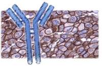
- ICC=1:50-200 IF=1:50-200
- HIV2
- HIV2

企业认证
相关产品推荐更多 >


万千商家帮你免费找货
0 人在求购买到急需产品
- 详细信息
- 文献和实验
- 技术资料
- 供应商:
上海联迈生物工程有限公司
- 库存:
大量
- 靶点:
详见说明书
- 级别:
1
- 目录编号:
LM-10482R-FITC
- 克隆性:
多克隆
- 抗原来源:
Rabbit
- 保质期:
1年
- 抗体英文名:
Anti-HIV2 gp120 + gp160/FITC
- 抗体名:
Anti-HIV2 gp120 + gp160/FITC
- 标记物:
FITC标记
- 宿主:
HIV2
- 适应物种:
HIV2
- 免疫原:
详见说明书
- 亚型:
IGg
- 形态:
粉末、液体、冻干粉
- 应用范围:
ICC=1:50-200 IF=1:50-200
- 浓度:
1mg/ml
- 保存条件:
-20 °C
- 规格:
100ul
| 英文名称 | Anti-HIV2 gp120 + gp160/FITC |
| 中文名称 | FITC标记的人类免疫缺陷病毒2型/2型艾滋病病毒gp41+gp160抗体 |
| 别 名 | Gp120; Gp160; HIV-2 gp120; HIV2 gp160; HIV2gp120; HIV2gp160; HIV 2; Human immunodeficiency virus 2; Human Immunodeficiency Virus Type 2; ENV_HV2RO. |
| 规格价格 | 100ul/2980元 购买 大包装/询价 |
| 说 明 书 | 100ul |
| 研究领域 | 细菌及病毒 |
| 抗体来源 | Rabbit |
| 克隆类型 | Polyclonal |
| 交叉反应 | HIV2 |
| 产品应用 | ICC=1:50-200 IF=1:50-200 not yet tested in other applications. optimal dilutions/concentrations should be determined by the end user. |
| 性 状 | Lyophilized or Liquid |
| 浓 度 | 1mg/ml |
| 免 疫 原 | KLH conjugated synthetic peptide derived from human HIV2 gp120 + gp160 |
| 亚 型 | IgG |
| 纯化方法 | affinity purified by Protein A |
| 储 存 液 | 0.01M TBS(pH7.4) with 1% BSA, 0.03% Proclin300 and 50% Glycerol. |
| 保存条件 | Store at -20 °C for one year. Avoid repeated freeze/thaw cycles. The lyophilized antibody is stable at room temperature for at least one month and for greater than a year when kept at -20°C. When reconstituted in sterile pH 7.4 0.01M PBS or diluent of antibody the antibody is stable for at least two weeks at 2-4 °C. |
| 产品介绍 | background: Human immunodeficiency virus type 2 (HIV2), orginally isolated from patients in West Africa, is the dominant form of HIV in West Africa capable of causing the acquired immunodeficiency syndrome (AIDS). HIV2 is closely related to simian immunodeficiency viruses (SIV). HIV1 and HIV2 share similarity in their genomes, transmission, clinical features, immunological effects, and in their action of binding to the same CD4 cellular receptor, but there are significant differences in the amino acid and nucleotide sequences of HIV1 and HIV2, especially within their envelope genes and proteins. Additionally, HIV2 may have a longer incubation period and may be less pathogenic than HIV1. HIV2 gp36 is a transmembrane protein located in the envelope of the virus specific to HIV2 that binds to the putative cellular receptor proteins P45 and P62. Function: The surface protein gp120 (SU) attaches the virus to the host lymphoid cell by binding to the primary receptor CD4. This interaction induces a structural rearrangement creating a high affinity binding site for a chemokine coreceptor like CXCR4 and/or CCR5. This peculiar 2 stage receptor-interaction strategy allows gp120 to maintain the highly conserved coreceptor-binding site in a cryptic conformation, protected from neutralizing antibodies. Since CD4 also displays a binding site for the disulfide-isomerase P4HB/PDI, a P4HB/PDI-CD4-CXCR4-gp120 complex may form. In that complex, P4HB/PDI could reach and reduce gp120 disulfide bonds, causing major conformational changes in gp120. TXN, another PDI family member could also be involved in disulfide rearrangements in Env during fusion. These changes are transmitted to the transmembrane protein gp41 and are thought to activate its fusogenic potential by unmasking its fusion peptide. The surface protein gp120 is a ligand for CD209/DC-SIGN and CLEC4M/DC-SIGNR, which are respectively found on dendritic cells (DCs), and on endothelial cells of liver sinusoids and lymph node sinuses. These interactions allow capture of viral particles at mucosal surfaces by these cells and subsequent transmission to permissive cells. DCs are professional antigen presenting cells, critical for host immunity by inducing specific immune responses against a broad variety of pathogens. They act as sentinels in various tissues where they take up antigen, process it, and present it to T-cells following migration to lymphoid organs. HIV subverts the migration properties of dendritic cells to gain access to CD4+ T-cells in lymph nodes. Virus transmission to permissive T-cells occurs either in trans (without DCs infection, through viral capture and transmission), or in cis (following DCs productive infection, through the usual CD4-gp120 interaction), thereby inducing a robust infection. In trans infection, bound virions remain infectious over days and it is proposed that they are not degraded, but protected in non-lysosomal acidic organelles within the DCs close to the cell membrane thus contributing to the viral infectious potential during DCs' migration from the periphery to the lymphoid tissues. On arrival at lymphoid tissues, intact virions recycle back to DCs' cell surface allowing virus transmission to CD4+ T-cells. Virion capture also seems to lead to MHC-II-restricted viral antigen presentation, and probably to the activation of HIV-specific CD4+ cells. The transmembrane protein gp41 (TM) acts as a class I viral fusion protein. Under the current model, the protein has at least 3 conformational states: pre-fusion native state, pre-hairpin intermediate state, and post-fusion hairpin state. During fusion of viral and target intracellular membranes, the coiled coil regions (heptad repeats) assume a trimer-of-hairpins structure, positioning the fusion peptide in close proximity to the C-terminal region of the ectodomain. The formation of this structure appears to drive apposition and subsequent fusion of viral and target cell membranes. Complete fusion occurs in host cell endosomes and is dynamin-dependent, however some lipid transfer might occur at the plasma membrane. The virus undergoes clathrin-dependent internalization long before endosomal fusion, thus minimizing the surface exposure of conserved viral epitopes during fusion and reducing the efficacy of inhibitors targeting these epitopes. Membranes fusion leads to delivery of the nucleocapsid into the cytoplasm. The envelope glyprotein gp160 precursor down-modulates cell surface CD4 antigen by interacting with it in the endoplasmic reticulum and blocking its transport to the cell surface. The gp120-gp41 heterodimer seems to contribute to T-cell depletion during HIV-1 infection. The envelope glycoproteins expressed on the surface of infected cells induce apoptosis through an interaction with uninfected cells expressing the receptor (CD4) and the coreceptors CXCR4 or CCR5. This type of bystander killing may be obtained by at least three distinct mechanisms. First, the interaction between the 2 cells can induce cellular fusion followed by nuclear fusion within the syncytium. Syncytia are condemned to die from apoptosis. Second, the 2 interacting cells may not fuse entirely and simply exchange plasma membrane lipids, after a sort of hemifusion process, followed by rapid death. Third, it is possible that virus-infected cells, on the point of undergoing apoptosis, fuse with CD4-expressing cells, in which case apoptosis is rapidly transmitted from one cell to the other and thus occurs in a sort of contagious fashion. The gp120-gp41 heterodimer allows rapid transcytosis of the virus through CD4 negative cells such as simple epithelial monolayers of the intestinal, rectal and endocervical epithelial barriers. Both gp120 and gp41 specifically recognize glycosphingolipids galactosyl-ceramide (GalCer) or 3' sulfo-galactosyl-ceramide (GalS) present in the lipid rafts structures of epithelial cells. Binding to these alternative receptors allows the rapid transcytosis of the virus through the epithelial cells. This transcytotic vesicle-mediated transport of virions from the apical side to the basolateral side of the epithelial cells does not involve infection of the cells themselves. Subunit: The mature envelope protein (Env) consists of a homotrimer of non-covalently associated gp120-gp41 heterodimers. The resulting complex protrudes from the virus surface as a spike. There seems to be as few as 10 spikes on the average virion. Surface protein gp120 interacts with human CD4, CCR5 and CXCR4, to form a P4HB/PDI-CD4-CXCR4-gp120 complex. Gp120 also interacts with the C-type lectins CD209/DC-SIGN and CLEC4M/DC-SIGNR (collectively referred to as DC-SIGN(R)). Gp120 and gp41 interact with GalCer. Subcellular Location: Transmembrane protein gp41: Virion membrane; Single-pass type I membrane protein. Host cell membrane; Single-pass type I membrane protein. Host endosome membrane; Single-pass type I membrane protein (Potential). Note=It is probably concentrated at the site of budding and incorporated into the virions possibly by contacts between the cytoplasmic tail of Env and the N-terminus of Gag. Surface protein gp120: Virion membrane; Peripheral membrane protein. Host cell membrane; Peripheral membrane protein. Host endosome membrane; Peripheral membrane protein (Potential). Note=The surface protein is not anchored to the viral envelope, but associates with the extravirion surface through its binding to TM. It is probably concentrated at the site of budding and incorporated into the virions possibly by contacts between the cytoplasmic tail of Env and the N-terminus of Gag. Post-translational modifications: Specific enzymatic cleavages in vivo yield mature proteins. Envelope glycoproteins are synthesized as a inactive precursor that is heavily N-glycosylated and processed likely by host cell furin in the Golgi to yield the mature SU and TM proteins. The cleavage site between SU and TM requires the minimal sequence [KR]-X-[KR]-R. Palmitoylation of the transmembrane protein and of Env polyprotein (prior to its proteolytic cleavage) is essential for their association with host cell membrane lipid rafts. Palmitoylation is therefore required for envelope trafficking to classical lipid rafts, but not for viral replication. Database links: UniProtKB/Swiss-Prot: P04577 Important Note: This product as supplied is intended for research use only, not for use in human, therapeutic or diagnostic applications. |
风险提示:丁香通仅作为第三方平台,为商家信息发布提供平台空间。用户咨询产品时请注意保护个人信息及财产安全,合理判断,谨慎选购商品,商家和用户对交易行为负责。对于医疗器械类产品,请先查证核实企业经营资质和医疗器械产品注册证情况。
 文献和实验
文献和实验抗人类免疫缺陷病毒(HIV)感染免疫 艾滋病(AIDS)由人类免疫缺陷病毒(HIV)感染导致,主要感染人体CD4T细胞,导致严重免疫缺陷。据联合国"2005年全球艾滋病传播报告”称:自1981年确认HIV病毒感染以来,迄今累计感染人口6 500万,已造成2 800万人死亡,现有AIDS患者3 800万;2005年死亡患者280万,新感染人口约400万;中国目前实际感染人数超过100万,进入快速增长期。由于AIDS导致的获得性免疫缺陷,患者通常伴有广谱性机会
艾滋病(AIDS)又称获得性免疫缺陷综合征,是由人类免疫缺陷病毒(HIV)感染所引起的一种免疫缺陷性疾病。HIV感染的诊断是艾滋病预防控制工作的重要组成部分,建立敏感实用的检测方法对于监测、诊断或血液筛查,控制艾滋病的流行显得尤为重要。以下本文就HIV的检测技术现状及进展做一综述。 1、HIV的感染机制及感染标志 HIV病毒侵入人体后,其表面糖蛋白gp120与细胞表面受体蛋白CD4以高亲和力结合,吸附到宿主细胞上;gp
HIV分型的分子流行病和临床意义: HIV在病毒分类中属逆转录病毒科慢病毒属中的人类免疫缺陷病毒组。由于以下4个方面的原因其基因有很高的变异特征,1.高错误率的逆转录酶,其复制时,不能及时切除错误引入的核苷酸,约每个复制循环中就会发生一个错误使病毒复制时发生随机变异。2.病毒快速的复制,估计每天一个HIV感染者要产生并清除100亿个病毒颗粒。3.宿主的免疫选择作用,在宿主免疫压力作用下,使能激发宿主细胞或体液免疫的基因组的变异高于其他部位,如gp120V3环高变区,4.不同病毒株DNA之间
 技术资料
技术资料暂无技术资料 索取技术资料

