


- ¥1500
- ATCC、DSMZ、ECACC、RIKEN
- 江苏
- CL1206
- 2026年05月29日

企业认证
相关产品推荐更多 >


万千商家帮你免费找货
0 人在求购买到急需产品
- 详细信息
- 文献和实验
- 技术资料
- 英文名:
YAC-1
- 库存:
100万
- 供应商:
欣润生物
- 肿瘤类型:
淋巴瘤
- 细胞类型:
细胞系
- ATCC Number:
详见说明
- 品系:
小鼠
- 组织来源:
淋巴
- 相关疾病:
淋巴瘤
- 物种来源:
小鼠
- 免疫类型:
不详
- 细胞形态:
淋巴母细胞样
- 是否是肿瘤细胞:
是
- 器官来源:
淋巴
- 运输方式:
新鲜或干冰
- 年限:
成年
- 生长状态:
悬浮生长
- 规格:
T25方瓶
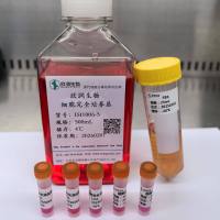
- 细胞名称:YAC-1细胞(小鼠淋巴瘤细胞)
- 形态:淋巴母细胞样,悬浮生长
- 含量:>1x10⁶ 个/瓶
- 污染:支原体、细菌、酵母和真菌检测为阴性
- 规格:T25瓶或者1mL冻存管包装
二、细胞接收后的处理:
1、贴壁细胞
- 收到T25方瓶细胞后,请检查是否漏液,如果漏液,请拍照片发给我们(冻存管细胞收到后直接37℃水浴复苏或直接放置于液氮中长期储存)。
- 请先在显微镜下确认细胞生长状态,去掉封口膜并将T25瓶置于37℃培养约2-3h。
- 弃去T25瓶中的培养基,换用新鲜的完全培养基。
- 如果细胞长满(90%以上)请及时进行细胞传代。
- 接到细胞次日,请检查细胞是否污染,若发现污染或疑似污染,请及时与我们取得联系。
2、悬浮细胞
- 收到细胞后,请检查是否漏液,如果漏液,请拍照片发给我们。
- 请先在显微镜下确认细胞生长状态,去掉封口膜并将15ml离心管置于37℃培养约2-3h。
- 1200rpm离心5min,弃去15ml离心管中的培养基,细胞沉淀用新鲜的完全培养基重悬并培养。
- 如果细胞长满(90%以上)请及时进行细胞传代。
- 接到细胞次日,请检查细胞是否污染,若发现污染或疑似污染,请及时与我们取得联系。
本公司的细胞培养操作规程,供参考
一、培养基及培养冻存条件准备:
- 准备RPMI-1640培养基,90%;优质胎牛血清,10%。
- 培养条件: 气相:空气,95%;二氧化碳,5%。 温度:37℃,培养箱湿度为70%-80%。
- 冻存液:90%血清,10%DMSO,现用现配。液氮储存。
对于贴壁细胞,传代可参考以下方法:
- 弃去培养上清,用不含钙、镁离子的PBS润洗细胞1-2次。
- 加2ml消化液(0.25%Trypsin-0.53mM EDTA)于培养瓶中,置于37℃培养箱中消化2-3分钟,然后在显微镜下观察细胞消化情况,若细胞大部分变圆并脱落,迅速拿回操作台,轻敲几下培养瓶后加入3ml此细胞的培养基终止消化。
- 轻轻吹打后吸出,移入15ml离心管中,在1200RPM条件下离心5分钟,弃去上清液,加入1mL培养液后吹匀。
- 移入到事先准备好的含有5ml培养基的T-25培养瓶中或含有14ml培养基的T-75培养瓶中培养。
3)细胞冻存:待细胞生长状态良好时,可进行细胞冻存。贴壁细胞冻存时,先要消化处理并进行细胞计数。消化方法按照细胞传代方法的1-3步骤进行,最后的重悬液使用血清。悬浮细胞直接计数后离心,用血清重悬浮,加DMSO至最终浓度为10%。加入DMSO后迅速混匀,按每1ml的数量分配到冻存管中。本公司按每个冻存管细胞数目大于1X10⁶个细胞冻存。
注意事项:
1. 收到冻存管细胞后,若发现干冰已挥发干净、冻存管瓶盖脱落、破损及细胞有污染,请立即与我们联系。
2. 所有动物细胞均视为有潜在的生物危害性,必须在二级生物安全台内操作,并请注意防护,所有废液及接触过此细胞的器皿需要灭菌后方能丢弃。
3. 细胞用途:仅供科研使用。
发货方式:
复苏后发货:我们复苏细胞后发货,货期一周左右,免运费。(气温较好建议复苏后发货)
冻存发货(干冰运输):需额外增加干冰运费,选择干冰运输的我们发两管细胞,为了保证客户接种可靠性多发一管。(气温低于0℃须冻存发货)
细胞发货采取专业的运输包装,并选择最快捷的运输方式(顺丰速运或其他空运快递)
Nonspecific cytotoxic cells of rainbow trout (Oncorhynchus mykiss) kill YAC-1 targets by both necrotic and apoptic mechanisms.
Nonspecific cytotoxic cells (NCC) have been identified in a number of fish species and are thought to be evolutionary progenitors of mammalian natural killer cells. We show here that trout NCC are functionally similar to cytotoxic cells of higher vertebrates in that they mediate cytotoxicity through both mechanisms of apoptosis and necrosis. To demonstrate that trout NCC inflict apoptic and necrotic lesions in tumor target cells, DNA fragmentation and 51chromium release assays were conducted using leukocytes isolated from peripheral blood, spleen, and anterior kidney. At effector-target ratios of 25:1, 50:1, 100:1, and 200:1, the release of thymidine-labeled DNA fragments and the release of 51chromium from YAC-1 target cells paralleled one another. Percent chromium release and DNA fragmentation increased when effector:target incubation times were extended from 4 to 18 h. As evidenced in agarose gels, the pattern of fragmentation induced by trout effector cells was identical to that produced by BALB/c NK cells. Similar to human and murine NK cells, trout NCC were maximally inhibited by 50 mM mannose-6-phosphate. Morphologic characteristics of rainbow trout NCC were examined using light and electron microscopy. Photomicrographs of effector:target cell mixtures after a 1 h incubation show NCC binding to target YAC-1 cells. Transmission electron micrographs of the conjugates revealed that the cells responsible for killing are small (4.2-4.5 microns), agranular mononuclear leukocytes.
Structural studies of gangliosides from the YAC-1 mouse lymphoma cell line by immunological detection and fast atom bombardment mass spectrometry
YAC-1 cells were propagated in bioreactors in 11 and 7.51 volumes. The cells were metabolically labelled with d -[1- 14 C]galactose and d -[1- 14 C]glucosamine. The ganglioside fraction, purified by DEAE-Sepharose and silica gel column chromatography, showed on thin layer chromatography four major bands with mobilities between G M1 and G D1a . Gangliosides, obtained by further purification steps including high performance liquid chromatography on silica gel 60 columns with a gradient system of isopropanol:hexane:water, and preparative high performance TLC were characterized by (1) immunostaining of corresponding asialogangliosides obtained by mild acid hydrolysis and neuraminidase treatment and (2) fast atom bombardment mass spectrometry of native and permethylated samples and methylation analysis of G M1b ganglioside. As well as small amounts of G M2 and G M1 , the major gangliosides found in the complex mixture were G M1b and GalNAc-G M1b . The structural heterogeneity of these gangliosides was cased by (a) substitution of the ceramide moiety by fatty acids of different chain length and degree of unsaturation (C 16:0 , C 24:0 , C 24:1 ) and (b) N-substitution of the sialic acid moieties with either acetyl or glycolyl groups. Disialogangliosides were detected only in low amounts and will be the subject of further investigation. A polyclonal chicken antiserum was raised against IVNeuAc-GgOse 5 Cer. The antiserum was highly specific for gangliosides (IVNeuAc and IVNeuGc) and asialogangliosides with a GgOse 5 Cer backbone. No cross-reaction with G M1b or GgOse 4 Cer was observed.
风险提示:丁香通仅作为第三方平台,为商家信息发布提供平台空间。用户咨询产品时请注意保护个人信息及财产安全,合理判断,谨慎选购商品,商家和用户对交易行为负责。对于医疗器械类产品,请先查证核实企业经营资质和医疗器械产品注册证情况。
 文献和实验
文献和实验Chicken intestinal epithelial cells were obtained from NEWGAINBIO company. Cells were cultured on 37℃, with 5% CO2, in the Ham’s F-12 Nutrient (DMEM/12) that contained the following supplementations: fetal bovine serum (5%), in-sulin (5 µg/mL), transferrin (5 µg/mL), selenium (5 ng/mL), epidermal growth factor (5 ng/mL) and penicillin-streptomycin (100–100 U/mL) for cell culturing (full DMEM/12). Experiments were performed with chicken intestinal epithelial cells and working solutions were prepared with plain DMEM/12 without supplementation. For the investigations, cells were seeded onto 96-well, 24-well or 6-well polystyrene cell culture plates.
Primary hVICs (passage 2) were cultured to 50–60% confluence and infected with pGMLV-SV40T-puro lentivirus (NewgainBio, Wuxi, China) at a multiplicity of infection of 80 supplemented with 5 µg/mL polybrene (Sigma-Aldrich, Buchs, Switzerland).
Tissue was cultured until cells became visible around the tissue, and when the fusion reached 90% (FIGURE 1A) §ask ¦lled with the prepared culturing medium was sent to the company for further immortalisation. Cell immortalisation was done for cell stability and longer-term use. Immortalised cells were cultured with 10% FBS and 1% PS in the DMEM medium. After the cells multiplied and merged, they were routinely passed and grown ( NEWGAINBIO Inc. Wuxi, Jiangsu, China) (FIGURE 1B-C).
Mouse primary cultured renal vascular ECs and VSMCs were obtained from Newgainbio company, which were tested by Factor VIII and α-smooth muscle actin (α-SMA), the marker of ECs and VSMCs. RNeasy Mini Kit was used for RNA extraction, and the above protocols were repeated.
Porcine primary colon epithelial cells (Newgainbio company, Wuxi,China) were cultured in Dulbecco's Modified Eagle's Medium (Solarbio, Beijing, China) containing 10 % fetal bovine serum (BioInd, Kiryat shmona, Lsrael) at 37 ◦C and 5 % CO2 humidity.
瘤/大鼠神经胶质细胞瘤杂交瘤细胞 P19 小鼠畸胎瘤细胞, 小鼠畸胎瘤细胞 PC-12 大鼠嗜铬细胞瘤细胞, 大鼠嗜铬细胞瘤 RAW264.7 小鼠巨噬细胞瘤 SP2/0-AG14 小鼠骨髓瘤 Yac-1 小鼠淋巴瘤细胞
):将外源目的DNA导入受体细胞,并能自我复制和增殖的工具。 载体具以下特征: 1)分子量小,便于携带较大的DNA片段,能进入宿主细胞并在其中增殖; 2)有多种限制酶切点,每种限制酶最好只有单一切点; 3)被切割后的载体,插入外源DNA后,不影响其复制能力,并有可选择的标记基因(如,抗药基因)。 常用的载体有:质粒,λ噬菌体,粘粒,BAC,YAC,PI等。(一).质粒plasmid Plasmid 独立于细菌染色体外的双链环DNA分子。 常用的质粒如pUC19,多连接子MCS。插入
Nature 子刊:迈出基因组重编码制备抗病毒人类细胞系的第一步
,初步证明了 TAG 转换为 TAA 在人类基因组中的可行性,同时创造了一次递送在人类基因组中数十个非重复位点同步碱基编辑的记录,为哺乳动物基因组的大规模工程化改造提供了一个工作框架。此外, GRIT 软件也可以被开发成一个新的计算机辅助设计 (CAD) 平台,用于设计编写大规模的基因组。 该研究迈出了基因组重编码制备抗多种天然病毒人类细胞系的第一步,为哺乳动物基因组多重复合编辑和 GP-write 路线图的制定奠定了基础。 虽然读取 DNA 密码的技术不断进步,但科学家们编写 DNA 密码


