


- ¥3500 - 4500
- 欣润生物
- 江苏无锡
- IH1019
- 2026年01月25日

企业认证
相关产品推荐更多 >


万千商家帮你免费找货
0 人在求购买到急需产品
- 详细信息
- 文献和实验
- 技术资料
- 英文名:
Human Umbilical Vein Smooth Muscle Cells
- 库存:
10
- 供应商:
欣润生物
- 肿瘤类型:
NO
- 细胞类型:
永生化
- ATCC Number:
11222
- 品系:
黄种
- 组织来源:
脐静脉平滑肌
- 相关疾病:
无
- 物种来源:
人源
- 免疫类型:
不详
- 细胞形态:
梭形
- 是否是肿瘤细胞:
否
- 器官来源:
/
- 运输方式:
常温
- 年限:
5年
- 生长状态:
贴壁生长
- 规格:
T25方瓶
永生化人脐静脉平滑肌细胞简介:
产品描述:脐静脉平滑肌细胞是从人脐静脉组织中提取的一种平滑肌细胞,具有贴壁生长的特性,常用于科研研究。脐静脉平滑肌细胞在血管病发过程中起重要作用,其异常增加的生长潜力在动脉疾病发生过程中具有决定性作用。这些细胞能够表达ICAM-1和VCAM-1,这些分子能促进血管壁中的炎症反应,并对血管疾病的发展和稳定起着重要作用。
产品货号:IH1019
产品类型: 原代细胞建立的永生化
传代能力: 30代左右
产品形态:梭形
培养基:永生化人脐静脉平滑肌细胞专用完全培养基,产品编号:IH1019-5
支原体:呈阴性
产品培养条件:37℃,5%CO2
发货方式:常温T25方瓶运输
货期:1周左右货期
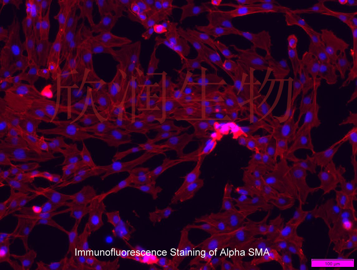
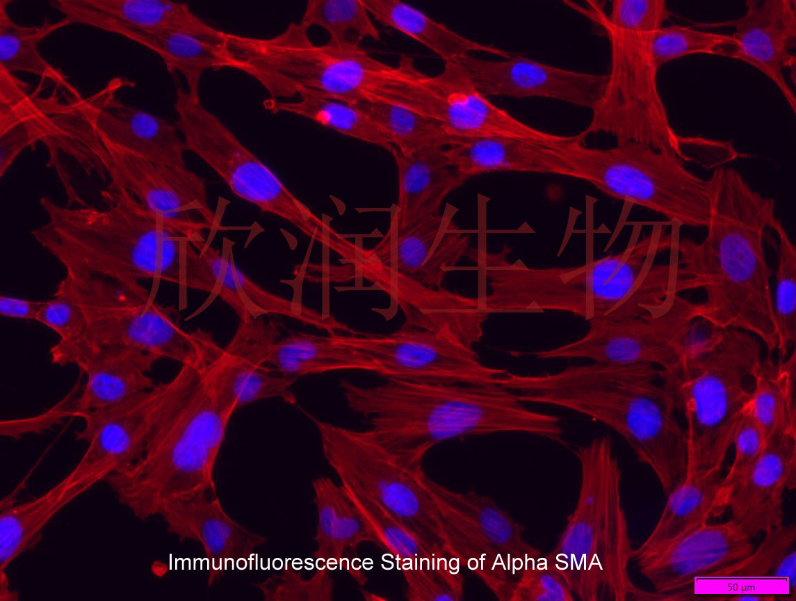
a-SMA抗体免疫荧光染色鉴定


Gp38k, a protein synthesized by vascular smooth muscle cells, stimulates directional migration of human umbilical vein endothelial cells
Gp38k is a 383-amino-acid secreted glycoprotein expressed by cultured vascular smooth muscle cells during the time of transition from a proliferating monolayer culture to a nonproliferating multilayered (differentiated) culture. Expression continues as the cell culture forms multicellular nodules. Because this transition period involves active cell migration, we evaluated the effects of exogenously added gp38k on vascular endothelial cell (HUVEC) migration and chemotaxis. Here we demonstrate that gp38k acts as a chemoattractant for HUVECs and stimulates cell migration in Boyden chambers at a level comparable to that achieved with the known endothelial cell chemoattractant bFGF. The migration effect is neutralized by the presence of a polyclonal anti-gp38k antibody. Because gp38k expression is also correlated with changes in culture morphology, we also assessed its ability to act as an agonist of HUVEC morphology using cultures growing on Matrigel. We report that gp38k stimulates endothelial cell tubulogenesis in this assay system. These results provide the first evidence
风险提示:丁香通仅作为第三方平台,为商家信息发布提供平台空间。用户咨询产品时请注意保护个人信息及财产安全,合理判断,谨慎选购商品,商家和用户对交易行为负责。对于医疗器械类产品,请先查证核实企业经营资质和医疗器械产品注册证情况。
 文献和实验
文献和实验Chicken intestinal epithelial cells were obtained from NEWGAINBIO company. Cells were cultured on 37℃, with 5% CO2, in the Ham’s F-12 Nutrient (DMEM/12) that contained the following supplementations: fetal bovine serum (5%), in-sulin (5 µg/mL), transferrin (5 µg/mL), selenium (5 ng/mL), epidermal growth factor (5 ng/mL) and penicillin-streptomycin (100–100 U/mL) for cell culturing (full DMEM/12). Experiments were performed with chicken intestinal epithelial cells and working solutions were prepared with plain DMEM/12 without supplementation. For the investigations, cells were seeded onto 96-well, 24-well or 6-well polystyrene cell culture plates.
Primary hVICs (passage 2) were cultured to 50–60% confluence and infected with pGMLV-SV40T-puro lentivirus (NewgainBio, Wuxi, China) at a multiplicity of infection of 80 supplemented with 5 µg/mL polybrene (Sigma-Aldrich, Buchs, Switzerland).
Tissue was cultured until cells became visible around the tissue, and when the fusion reached 90% (FIGURE 1A) §ask ¦lled with the prepared culturing medium was sent to the company for further immortalisation. Cell immortalisation was done for cell stability and longer-term use. Immortalised cells were cultured with 10% FBS and 1% PS in the DMEM medium. After the cells multiplied and merged, they were routinely passed and grown ( NEWGAINBIO Inc. Wuxi, Jiangsu, China) (FIGURE 1B-C).
Mouse primary cultured renal vascular ECs and VSMCs were obtained from Newgainbio company, which were tested by Factor VIII and α-smooth muscle actin (α-SMA), the marker of ECs and VSMCs. RNeasy Mini Kit was used for RNA extraction, and the above protocols were repeated.
Porcine primary colon epithelial cells (Newgainbio company, Wuxi,China) were cultured in Dulbecco's Modified Eagle's Medium (Solarbio, Beijing, China) containing 10 % fetal bovine serum (BioInd, Kiryat shmona, Lsrael) at 37 ◦C and 5 % CO2 humidity.
 技术资料
技术资料暂无技术资料 索取技术资料

